Human/Mouse Caspase-3 Antibody Summary
Met1-His277
Accession # AAA65015
Applications
Please Note: Optimal dilutions should be determined by each laboratory for each application. General Protocols are available in the Technical Information section on our website.
Scientific Data
 View Larger
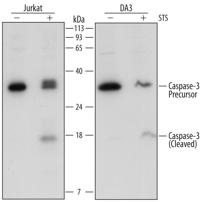
Detection of Human and Mouse Caspase‑3 by Western Blot. Western blot shows lysates of Jurkat human acute T cell leukemia cell line and DA3 mouse myeloma cell line untreated (-) or treated (+) with 1 µg/mL Staurosporine (STS) for 12 hours. PVDF Membrane was probed with 0.5 µg/mL of Goat Anti-Human Caspase-3 Antigen Affinity-purified Polyclonal Antibody (Catalog # AF-605-NA) followed by HRP-conjugated Anti-Goat IgG Secondary Antibody (Catalog # HAF017). Specific bands were detected for Caspase-3 precursor at approximately 34 kDa (as indicated) and Caspase-3 (cleaved) at approximately 18 kDa (as indicated). This experiment was conducted under reducing conditions and using Immunoblot Buffer Group 2.
 View Larger
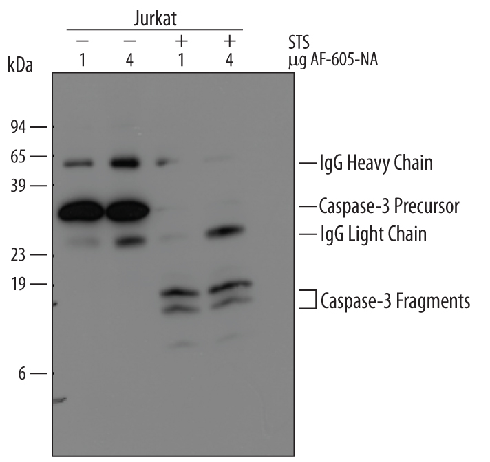
Immunoprecipitation of Human Caspase‑3. Jurkat human acute T cell leukemia cell line was treated with indicated concentrations of Staurosporine for 0 or 4 hours. Caspase‑3 was immunoprecipitated from lysates of 1‑2 x 10⁶cells following incubation with 1 or 4 µg Goat Anti-Human Caspase‑3 Antigen Affinity-purified Polyclonal Antibody (Catalog # AF-605-NA) overnight at 4 ºC. Caspase‑3-antibody complexes were absorbed using Protein G expressing Staph cells (Sigma). Immunoprecipitated Caspase‑3 was detected by Western blot using 0.5 µg/mL Goat Anti-Human Caspase‑3 Antigen Affinity-purified Polyclonal Antibody (Catalog # AF‑605‑NA). View our recommended buffer recipes for immunoprecipitation.
 View Larger
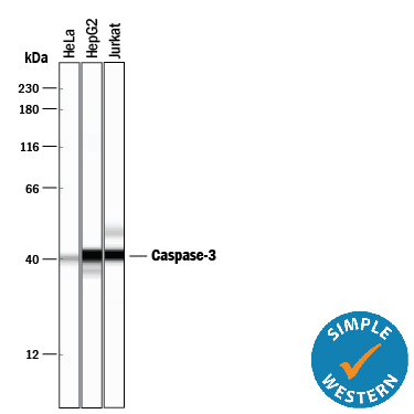
Detection of Human Caspase‑3 by Simple WesternTM. Simple Western lane view shows lysates of HeLa human cervical epithelial carcinoma cell line, HepG2 human hepatocellular carcinoma cell line, and Jurkat human acute T cell leukemia cell line, loaded at 0.2 mg/mL. A specific band was detected for Caspase-3 at approximately 40 kDa (as indicated) using 5 µg/mL of Goat Anti-Human/Mouse Caspase-3 Antigen Affinity-purified Polyclonal Antibody (Catalog # AF-605-NA) 1:50 dilution followed by HRP-conjugated Anti-Goat IgG Secondary Antibody (Catalog # HAF017). This experiment was conducted under reducing conditions and using the 12-230 kDa separation system.
 View Larger
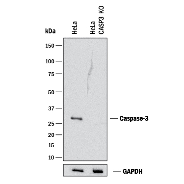
Western Blot Shows Human Caspase‑3 Specificity by Using Knockout Cell Line. Western blot shows lysates of HeLa human cervical epithelial carcinoma parental cell line and Caspase-3 knockout HeLa cell line (KO). PVDF membrane was probed with 0.2 µg/mL of Goat Anti-Human/Mouse Caspase-3 Antigen Affinity-purified Polyclonal Antibody (Catalog # AF-605-NA) followed by HRP-conjugated Anti-Goat IgG Secondary Antibody (Catalog # HAF017). A specific band was detected for Caspase-3 at approximately 32 kDa (as indicated) in the parental HeLa cell line, but is not detectable in knockout HeLa cell line. GAPDH (Catalog # AF5718) is shown as a loading control. This experiment was conducted under reducing conditions and using Immunoblot Buffer Group 1.
Reconstitution Calculator
Preparation and Storage
- 12 months from date of receipt, -20 to -70 °C as supplied.
- 1 month, 2 to 8 °C under sterile conditions after reconstitution.
- 6 months, -20 to -70 °C under sterile conditions after reconstitution.
Background: Caspase-3
Caspase-3 (Cysteine-aspartic acid protease 3/Casp3; also Yama, apopain and CPP32) is a 29 kDa member of the peptidase C14A family of enzymes (1, 2, 3). It is widely expressed and is an integral component of the apoptotic cascade. Caspase-3 is considered to be the major executioner caspase; that is, the primary downstream mediator of apoptotic-associated proteolysis (2, 3, 4). Active Caspase-3 is known to utilize a Cys residue to cleave multiple substrates, including PARP, proIL-16, PKC-gamma & -δ, procaspases 6, 7 and 9, and beta -catenin (1). Human procaspase-3 is a 32 kDa, 277 amino acid (aa) protein (5, 6, 7). Normally, it is an inactive, cytosolic homodimer, but following an upstream signal that activates processing proteases, procaspase-3 undergoes proteolytic cleavage (1, 2, 8, 9). This generates an N-terminal 175 aa p20/20 kDa subunit plus a 102 aa C-terminal p12/12 kDa subunit, followed by further processing of the p20 subunit at Asp28 to generate a final p17 subunit (aa 29-175) (9). The p17 and p12 subunits noncovalently heterodimerize, and subsequently associate with another p17/p12 heterodimer to form an active antiparallel homodimer. The p17 subunit contains the enzyme active site (aa 161-165), with an embedded catalytic Cys which is normally nitrosylated and inactive. Full activation requires both proteolytic processing and Cys163 denitrosylation (10). Multiple proteases can use Caspase-3 as a substrate including Caspase-6, -8, and -10, granzyme B, and Caspase-3 itself (9, 11, 12, 13).
-
Chowdhury, I. et al. (2008) Comp. Biochem. Physiol. B 151:10.
-
Boatright, K.M. & G.S. Salvesen (2003) Curr. Opin. Cell Biol. 15:725.
-
Launay, S. et al. (2005) Oncogene 24:5137.
-
Walsh, J.G. et al. (2008) Proc. Natl. Scad. Sci. USA 105:12815.
-
Nicholson, D.W. et al. (1995) Nature 376:37.
-
Tewari, M. et al. (1995) Cell 81:801.
-
Fernandes-Alnemri, T. et al. (1994) J. Biol. Chem. 269:30761.
-
Milisav, I. et al. (2009) Apoptosis 14:1070.
-
Han, Z. et al. (1997) J. Biol. Chem. 272:13432.
-
Rossig, L. et al. (1999) J. Biol. Chem. 274:6823.
-
Rank, K.B. et al. (2001) Protein Expr. Purif. 22:258.
-
Atkinson, E.A. et al. (1998) J. Biol. Chem. 273:21261.
- Cohen, G.M. (1997) Biochem. J. 326:1.
Product Datasheets
Citations for Human/Mouse Caspase-3 Antibody
R&D Systems personnel manually curate a database that contains references using R&D Systems products. The data collected includes not only links to publications in PubMed, but also provides information about sample types, species, and experimental conditions.
13
Citations: Showing 1 - 10
Filter your results:
Filter by:
-
Enhanced Apoptosis and Loss of Cell Viability in Melanoma Cells by Combined Inhibition of ERK and Mcl-1 Is Related to Loss of Mitochondrial Membrane Potential, Caspase Activation and Upregulation of Proapoptotic Bcl-2 Proteins
Authors: Z Peng, B Gillissen, A Richter, T Sinnberg, MS Schlaak, J Eberle
International Journal of Molecular Sciences, 2023-03-04;24(5):.
Species: Human
Sample Types: Cell Lysates
Applications: Western Blot -
Toxicity Assessment of Curculigo orchioides Leaf Extract Using Drosophila melanogaster: A Preliminary Study
Authors: S Kushalan, LC D'Souza, K Aloysius, A Sharma, S Hegde
International Journal of Environmental Research and Public Health, 2022-11-18;19(22):.
Species: Drosophila melanogaster
Sample Types: Tissue Homogenates
Applications: IHC -
Activation of the executioner caspases-3 and -7 promotes microglial pyroptosis in models of multiple sclerosis
Authors: BA McKenzie, JP Fernandes, MAL Doan, LM Schmitt, WG Branton, C Power
J Neuroinflammation, 2020-08-29;17(1):253.
Species: Human
Sample Types: Protein, Tissue Homogenates, Whole Cells, Whole Tissue
Applications: ICC, IHC, Western Blot -
Hypoxia-induced complement dysregulation is associated with microvascular impairments in mouse tracheal transplants
Authors: MA Khan, T Shamma, S Kazmi, A Altuhami, HA Ahmed, AM Assiri, DC Broering
J Transl Med, 2020-03-31;18(1):147.
Species: Mouse
Sample Types: Whole Tissue
Applications: IHC -
Apoptosis-mediated antiproliferation of A549 lung cancer cells mediated by Eugenia aquea leaf compound 2',4'-dihydroxy-6'-methoxy-3',5'-dimethylchalcone and its molecular interaction with caspase receptor in molecular docking simulation
Authors: YE Hadisaputr, N Cahyana, M Muchtaridi, R Lesmana, T Rusdiana, AY Chaerunisa, I Sufiawati, T Rostinawat, A Subarnas
Oncol Lett, 2020-03-19;19(5):3551-3557.
Species: Human
Sample Types: Cell Lysate
Applications: Western Blot -
Levels of caspase-3 and histidine-rich glycoprotein in the embryo secretome as biomarkers of good-quality day-2 embryos and high-quality blastocysts
Authors: H Kaihola, FG Yaldir, T Bohlin, R Samir, J Hreinsson, H Åkerud
PLoS ONE, 2019-12-19;14(12):e0226419.
Species: Human
Sample Types: Tissue Culture Supernates
Applications: Proximity Extension Assay -
Lipid storage droplet protein 5 reduces sodium palmitate?induced lipotoxicity in human normal liver cells by regulating lipid metabolism?related factors
Authors: X Ma, F Cheng, K Yuan, K Jiang, T Zhu
Mol Med Rep, 2019-06-06;20(2):879-886.
Species: Human
Sample Types: Cell Lysates
Applications: Western Blot -
The Effect of Long-Term Administration of Fatty Acid Amide Hydrolase Inhibitor URB597 on Oxidative Metabolism in the Heart of Rats with Primary and Secondary Hypertension
Authors: M Biernacki, W ?uczaj, I Jarocka-Ka, E Ambro?ewic, M Toczek, E Skrzydlews
Molecules, 2018-09-14;23(9):.
Species: Rat
Sample Types: Tissue Homogenates
Applications: Western Blot -
Bok is a genuine multi-BH-domain protein that triggers apoptosis in the absence of Bax and Bak and augments drug response
Authors: S Einsele-Sc, S Malmsheime, K Bertram, D Stehle, J Johänning, M Manz, PT Daniel, BF Gillissen, K Schulze-Os, F Essmann
J Cell Sci, 2016-04-13;0(0):.
Species: Human
Sample Types: Cell Lysates
Applications: Western Blot -
Overexpressing cellular repressor of E1A-stimulated genes protects mesenchymal stem cells against hypoxia- and serum deprivation-induced apoptosis by activation of PI3K/Akt.
Authors: Deng J, Han Y, Yan C, Tian X, Tao J, Kang J, Li S
Apoptosis, 2010-04-01;15(4):463-73.
Species: Rat
Sample Types: Cell Lysates
Applications: Western Blot -
Ellipticine derivative NSC 338258 represents a potential new antineoplastic agent for the treatment of multiple myeloma.
Authors: Tian E, Landowski TH, Stephens OW, Yaccoby S, Barlogie B, Shaughnessy JD
Mol. Cancer Ther., 2008-03-04;7(3):500-9.
Species: Human
Sample Types: Cell Lysates
Applications: Western Blot -
Photochemically mediated delivery of AdhCMV-TRAIL augments the TRAIL-induced apoptosis in colorectal cancer cell lines.
Authors: Engesaeter BØ, Bonsted A, Lillehammer T, Engebraaten O, Berg K, Maelandsmo GM
Cancer Biol. Ther., 2006-11-19;5(11):1511-20.
Species: Human
Sample Types: Cell Lysates
Applications: Western Blot -
The B30.2 domain of pyrin, the familial Mediterranean fever protein, interacts directly with caspase-1 to modulate IL-1beta production.
Authors: Chae JJ, Wood G, Masters SL, Richard K, Park G, Smith BJ, Kastner DL
Proc. Natl. Acad. Sci. U.S.A., 2006-06-19;103(26):9982-7.
Species: Human
Sample Types: Cell Lysates
Applications: Western Blot
FAQs
No product specific FAQs exist for this product, however you may
View all Antibody FAQsReviews for Human/Mouse Caspase-3 Antibody
Average Rating: 4 (Based on 1 Review)
Have you used Human/Mouse Caspase-3 Antibody?
Submit a review and receive an Amazon gift card.
$25/€18/£15/$25CAN/¥75 Yuan/¥2500 Yen for a review with an image
$10/€7/£6/$10 CAD/¥70 Yuan/¥1110 Yen for a review without an image
Filter by:


