Human/Mouse/Rat Actin Antibody Summary
Applications
Please Note: Optimal dilutions should be determined by each laboratory for each application. General Protocols are available in the Technical Information section on our website.
Scientific Data
 View Larger
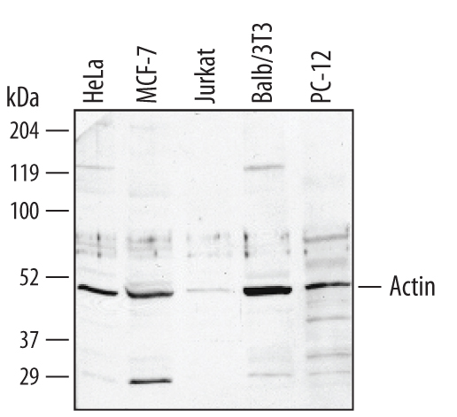
Detection of Human/Mouse/Rat Actin by Western Blot. Western blot shows lysates of HeLa human cervical epithelial carcinoma cell line, MCF-7 human breast cancer cell line, Jurkat human acute T cell leukemia cell line, Balb/3T3 mouse embryonic fibroblast cell line, and PC-12 rat adrenal pheochromocytoma cell line. PVDF membrane was probed with 1 µg/mL of Sheep Anti-Human/Mouse/Rat Actin Antigen Affinity-purified Polyclonal Antibody (Catalog # AF4000) followed by HRP-conjugated Anti-Sheep IgG Secondary Antibody (Catalog # HAF016). A specific band was detected for Actin at approximately 50 kDa (as indicated). This experiment was conducted under reducing conditions and using Immunoblot Buffer Group 1.
 View Larger
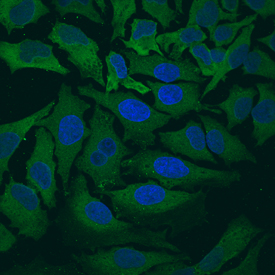
Actin in HeLa Human Cell Line. Actin was detected in immersion fixed HeLa human cervical epithelial carcinoma cell line using Sheep Anti-Human/Mouse/Rat Actin Antigen Affinity-purified Polyclonal Antibody (Catalog # AF4000) at 10 µg/mL for 3 hours at room temperature. Cells were stained using the NorthernLights™ 493-conjugated Anti-Sheep IgG Secondary Antibody (green; Catalog # NL012) and counterstained with DAPI (blue). View our protocol for Fluorescent ICC Staining of Cells on Coverslips.
 View Larger
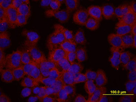
Actin in HEK293 Human Cell Line. Actin was detected in immersion fixed HEK293 human embryonic kidney cell line using Sheep Anti-Human/Mouse/Rat Actin Antigen Affinity-purified Polyclonal Antibody (Catalog # AF4000) at 10 µg/mL for 3 hours at room temperature. Cells were stained using the NorthernLights™ 557-conjugated Anti-Sheep IgG Secondary Antibody (red; Catalog # NL010) and counterstained with DAPI (blue). View our protocol for Fluorescent ICC Staining of Cells on Coverslips.
Reconstitution Calculator
Preparation and Storage
- 12 months from date of receipt, -20 to -70 °C as supplied.
- 1 month, 2 to 8 °C under sterile conditions after reconstitution.
- 6 months, -20 to -70 °C under sterile conditions after reconstitution.
Background: Actin
The Actin protein, a component of the eukaryotic cytoskeleton, is highly conserved between multiple species. Levels of the Actin protein may vary between different cell types, but are consistent within a given cell type, and for this reason, Actin protein levels are considered an internal control for protein loading.
Product Datasheets
Citations for Human/Mouse/Rat Actin Antibody
R&D Systems personnel manually curate a database that contains references using R&D Systems products. The data collected includes not only links to publications in PubMed, but also provides information about sample types, species, and experimental conditions.
5
Citations: Showing 1 - 5
Filter your results:
Filter by:
-
Fatty acid-binding protein 5 (FABP5) regulates cognitive function both by decreasing anandamide levels and by activating the nuclear receptor peroxisome proliferator-activated receptor beta/delta (PPARbeta/delta) in the brain.
Authors: Yu S, Levi L, Casadesus G, Kunos G, Noy N
J Biol Chem, 2014-03-18;289(18):12748-58.
Species: Mouse
Sample Types: Cell Lysates
Applications: Western Blot -
Nuclear trafficking of the HIV-1 pre-integration complex depends on the ADAM10 intracellular domain.
Authors: Endsley M, Somasunderam A, Li G, Oezguen N, Thiviyanathan V, Murray J, Rubin D, Hodge T, O'Brien W, Lewis B, Ferguson M
Virology, 2014-02-22;454(0):60-6.
Species: Human
Sample Types: Whole Cells
Applications: Western Blot Control -
Microbial exposure early in life regulates airway inflammation in mice after infection with Streptococcus pneumoniae with enhancement of local resistance.
Authors: Yasuda Y, Matsumura Y, Kasahara K, Ouji N, Sugiura S, Mikasa K, Kita E
Am. J. Physiol. Lung Cell Mol. Physiol., 2009-09-25;298(1):L67-78.
Species: Mouse
Sample Types: Tissue Homogenates
Applications: Western Blot -
cAMP/PKA pathway activation in human mesenchymal stem cells in vitro results in robust bone formation in vivo.
Authors: Siddappa R, Martens A, Doorn J, Leusink A, Olivo C, Licht R, van Rijn L, Gaspar C, Fodde R, Janssen F, van Blitterswijk C, de Boer J
Proc. Natl. Acad. Sci. U.S.A., 2008-05-19;105(20):7281-6.
Species: Human
Sample Types: Cell Lysates
Applications: Western Blot -
Accelerated formation of multicellular 3-D structures by cell-to-cell cross-linking.
Authors: De Bank PA, Hou Q, Warner RM, Wood IV, Ali BE, Macneil S, Kendall DA, Kellam B, Shakesheff KM, Buttery LD
Biotechnol. Bioeng., 2007-08-15;97(6):1617-25.
Species: Human
Sample Types: Cell Lysates
Applications: Western Blot
FAQs
No product specific FAQs exist for this product, however you may
View all Antibody FAQsReviews for Human/Mouse/Rat Actin Antibody
There are currently no reviews for this product. Be the first to review Human/Mouse/Rat Actin Antibody and earn rewards!
Have you used Human/Mouse/Rat Actin Antibody?
Submit a review and receive an Amazon gift card.
$25/€18/£15/$25CAN/¥75 Yuan/¥2500 Yen for a review with an image
$10/€7/£6/$10 CAD/¥70 Yuan/¥1110 Yen for a review without an image

