Human/Mouse/Rat Akt Pan Specific Antibody Summary
Ser2-Ala480
Accession # P31749
Applications
Please Note: Optimal dilutions should be determined by each laboratory for each application. General Protocols are available in the Technical Information section on our website.
Scientific Data
 View Larger
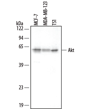
Detection of Human/Mouse Akt by Western Blot. Western blot shows lysates of MCF-7 human breast cancer cell line, MBA-MB-123 human breast cancer cell line, and TS1 mouse helper T cell line. PVDF membrane was probed with 0.2 µg/mL Mouse Anti-Human/Mouse/Rat Akt Pan Specific Monoclonal Antibody (Catalog # MAB2055) followed by HRP-conjugated Anti-Mouse IgG Secondary Antibody (Catalog # HAF007). This experiment was conducted under reducing conditions and using Immunoblot Buffer Group 4.
 View Larger
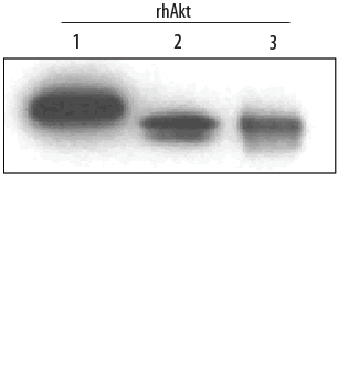
Detection of Human Akt Pan Specific by Western Blot. Western blot shows Recombinant Human Active Akt1 (Catalog # 1775-KS), recombinant human Akt2, and recombinant human Akt3 (5 ng/lane). PVDF membrane was probed with 0.2 µg/mL Mouse Anti-Human/Mouse/Rat Akt Pan Specific Monoclonal Antibody (Catalog # MAB2055) followed by HRP-conjugated Anti-Mouse IgG Secondary Antibody (Catalog # HAF007). This experiment was conducted under reducing conditions and using Immunoblot Buffer Group 4.
 View Larger
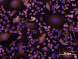
Akt in MDA‑MB‑231 Human Cell Line. Akt was detected in immersion fixed MDA-MB-231 human breast cancer cell line using Mouse Anti-Human/ Mouse/Rat Akt Monoclonal Antibody (Catalog # MAB2055) at 10 µg/mL for 3 hours at room temperature. Cells were stained using the NorthernLights™ 557-conjugated Anti-Mouse IgG Secondary Antibody (yellow; Catalog # NL007) and counter-stained with DAPI (blue). View our protocol for Fluorescent ICC Staining of Cells on Coverslips.
 View Larger
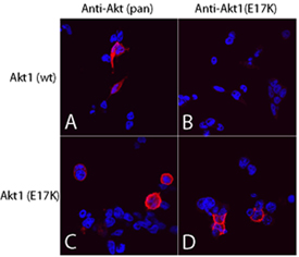
Akt (pan) in 293T Human Cell Line. Akt pan specific (panels A and C) and Akt1 (E17K Mutation) (panels B and D) were detected in immersion fixed 293T human embryonic kidney cell line transfected with wild type (panels A and B) or E17K mutated (panels C and D) Akt1 using Mouse Anti-Human/Mouse/Rat Akt Pan Specific Monoclonal Antibody (Catalog # MAB2055) and Mouse Anti-Human Akt1 (E17K Mutation) Monoclonal Antibody (Catalog # MAB6815). Both antibodies were used at 10 µg/mL for 3 hours at room temperature. Cells were stained using the NorthernLights™ 557-conjugated Anti-Mouse IgG Secondary Antibody (red; Catalog # NL007) and counterstained with DAPI (blue). Specific staining was localized to plasma membranes and cytoplasm. View our protocol for Fluorescent ICC Staining of Cells on Coverslips.
 View Larger
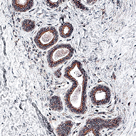
Akt Pan Specific in Human Breast Cancer Tissue. Akt was detected in immersion fixed paraffin-embedded sections of human breast cancer tissue using Mouse Anti-Human/Mouse/Rat Akt Pan Specific Monoclonal Antibody (Catalog # MAB2055) at 5 µg/mL for 1 hour at room temperature followed by incubation with the Anti-Mouse IgG VisUCyte™ HRP Polymer Antibody (VC001). Before incubation with the primary antibody, tissue was subjected to heat-induced epitope retrieval using Antigen Retrieval Reagent-Basic (CTS013). Tissue was stained using DAB (brown) and counterstained with hematoxylin (blue). Specific staining was localized to cytoplasm in epithelial cells. Staining was performed using our IHC Staining with VisUCyte HRP Polymer Detection Reagents.
 View Larger
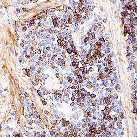
Akt Pan Specific in Mouse Spleen Tissue. Akt was detected in immersion fixed paraffin-embedded sections of mouse spleen tissue using Mouse Anti-Human/Mouse/Rat Akt Pan Specific Monoclonal Antibody (Catalog # MAB2055) at 5 µg/mL for 1 hour at room temperature followed by incubation with the Anti-Mouse IgG VisUCyte™ HRP Polymer Antibody (VC001). Before incubation with the primary antibody, tissue was subjected to heat-induced epitope retrieval using Antigen Retrieval Reagent-Basic (CTS013). Tissue was stained using DAB (brown) and counterstained with hematoxylin (blue). Specific staining was localized to cytoplasm in lymphocytes. Staining was performed using our IHC Staining with VisUCyte HRP Polymer Detection Reagents.
 View Larger
Detection of Akt in MCF‑7 Human Cell Line by Flow Cytometry. MCF-7 human breast cancer cell line was stained with Mouse Anti-Human/Mouse/Rat Akt Monoclonal Antibody (Catalog # MAB2055, filled histogram) or isotype control antibody (Catalog # MAB0041, open histogram), followed by Phycoerythrin-conjugated Anti-Mouse IgG F(ab')2Secondary Antibody (Catalog # F0102B). To facilitate intracellular staining, cells were fixed with paraformaldehyde and permeabilized with saponin.
 View Larger
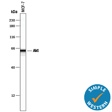
Detection of Human Akt by Simple WesternTM. Simple Western lane view shows lysates of MCF‑7 human breast cancer cell line, loaded at 0.2 mg/mL. A specific band was detected for Akt at approximately 62 kDa (as indicated) using 2 µg/mL of Mouse Anti-Human/Mouse/Rat Akt Pan Specific Monoclonal Antibody (Catalog # MAB2055). This experiment was conducted under reducing conditions and using the 12-230 kDa separation system.
Reconstitution Calculator
Preparation and Storage
- 12 months from date of receipt, -20 to -70 °C as supplied.
- 1 month, 2 to 8 °C under sterile conditions after reconstitution.
- 6 months, -20 to -70 °C under sterile conditions after reconstitution.
Background: Akt
Akt, also known as protein kinase B (PKB), is a central kinase in such diverse cellular processes as glucose uptake, cell cycle progression, and apoptosis. Three highly homologous members define the Akt family: Akt1 (PKB alpha ), Akt2 (PKB beta ), and Akt3 (PKB gamma ). All three Akts contain an amino-terminal pleckstrin homology domain, a central kinase domain, and a carboxyl-terminal regulatory domain.
Product Datasheets
Citations for Human/Mouse/Rat Akt Pan Specific Antibody
R&D Systems personnel manually curate a database that contains references using R&D Systems products. The data collected includes not only links to publications in PubMed, but also provides information about sample types, species, and experimental conditions.
18
Citations: Showing 1 - 10
Filter your results:
Filter by:
-
Approaches in Hydroxytyrosol Supplementation on Epithelial-Mesenchymal Transition in TGFbeta1-Induced Human Respiratory Epithelial Cells
Authors: RA Razali, MD Yazid, A Saim, RBH Idrus, Y Lokanathan
International Journal of Molecular Sciences, 2023-02-16;24(4):.
Species: Human
Sample Types: Cell Lysates
Applications: Western Blot -
A Three-Dimensional Xeno-Free Culture Condition for Wharton's Jelly-Mesenchymal Stem Cells: The Pros and Cons
Authors: B Koh, N Sulaiman, MB Fauzi, JX Law, MH Ng, TL Yuan, AGN Azurah, MH Mohd Yunus, RBH Idrus, MD Yazid
International Journal of Molecular Sciences, 2023-02-13;24(4):.
Species: Human
Sample Types: Cell Lysates
Applications: Western Blot -
Dock7 regulates AKT and mTOR/S6K activity required for the transformed phenotypes and survival of cancer cells
Authors: OY Teran, MR Zanotelli, MJ Lin, RA Cerione, KF Wilson
bioRxiv : the preprint server for biology, 2023-01-03;0(0):.
Species: N/A
Sample Types: Recombinant Protein
Applications: Bioassay -
The proprotein convertase furin regulates the development of thymic epithelial cells to ensure central immune tolerance
Authors: Liang Z, Zhang Z, Zhang Q et al.
iScience
-
LncCDH5-3:3 Regulates Apoptosis, Proliferation, and Aggressiveness in Human Lung Cancer Cells
Authors: K Kwa?niak, J Czarnik-Kw, K Malysheva, K Pogoda, O Korchynsky, P Rybojad, B Karczmarek, J Tabarkiewi
Cells, 2022-01-23;11(3):.
Species: Human
Sample Types: Cell Lysates
Applications: Western Blot -
A growth-factor-activated lysosomal K+ channel regulates Parkinson's pathology
Authors: J Wie, Z Liu, H Song, TF Tropea, L Yang, H Wang, Y Liang, C Cang, K Aranda, J Lohmann, J Yang, B Lu, AS Chen-Plotk, KC Luk, D Ren
Nature, 2021-01-27;0(0):.
Species: Human, Mouse, Transgenic Mouse
Sample Types: Cell Lysates
Applications: Western Blot -
ACPA decreases non-small cell lung cancer line growth through Akt/PI3K and JNK pathways in vitro
Authors: Ö Boyac?o?lu, E Bilgiç, C Varan, E Bilensoy, E Nemutlu, D Sevim, Ç Kocaefe, P Korkusuz
Cell Death & Disease, 2021-01-11;12(1):56.
Species: Human
Sample Types: Cell Lysates
Applications: Western Blot -
Multiplex coherent anti-Stokes Raman scattering microspectroscopy detection of lipid droplets in cancer cells expressing TrkB
Authors: T Guerenne-D, V Couderc, L Duponchel, V Sol, P Leproux, JM Petit
Sci Rep, 2020-10-07;10(1):16749.
Species: Human
Sample Types: Cell Lysates
Applications: Western Blot -
Jejunal Insulin Signalling Is Increased in Morbidly Obese Subjects with High Insulin Resistance and Is Regulated by Insulin and Leptin
Authors: C Gutierrez-, A Ho-Plagaro, C Santiago-F, S Garcia-Ser, F Rodríguez-, S Valdes, L Garrido-Sa, C Rodríguez-, C López-Góme, FJ Moreno-Rui, G Alcain-Mar, A Gautier-St, G Mithieux, E Garcia-Fue
J Clin Med, 2020-01-10;9(1):.
Species: Human
Sample Types: Tissue Homogenates
Applications: Western Blot -
C16 Peptide Promotes Vascular Growth and Reduces Inflammation in a Neuromyelitis Optica Model
Authors: H Chen, X Fu, J Jiang, S Han
Front Pharmacol, 2019-12-03;10(0):1373.
Species: Rat
Sample Types: Whole Tissue
Applications: IHC -
Subthalamic Nucleus Deep Brain Stimulation Employs trkB Signaling for Neuroprotection and Functional Restoration
Authors: DL Fischer, CJ Kemp, A Cole-Strau, NK Polinski, KL Paumier, JW Lipton, K Steece-Col, TJ Collier, DJ Buhlinger, CE Sortwell
J. Neurosci., 2017-06-12;37(28):6786-6796.
Species: Mouse
Sample Types: Whole Tissue
Applications: IHC-Fr -
Adipocyte SIRT1 controls systemic insulin sensitivity by modulating macrophages in adipose tissue
Authors: X Hui, M Zhang, P Gu, K Li, Y Gao, D Wu, Y Wang, A Xu
EMBO Rep, 2017-03-07;0(0):.
Species: Mouse
Sample Types: Tissue Homogenates
Applications: Western Blot -
Endocrine responses and acute mTOR pathway phosphorylation to resistance exercise with leucine and whey
Authors: MT Lane, TJ Herda, AC Fry, MA Cooper, MJ Andre, PM Gallagher
Biol Sport, 2017-01-20;34(2):197-203.
Species: Human
Sample Types: Tissue Homogenates
Applications: Infared Microscopy, Western Blot -
Lack of CD2AP disrupts Glut4 trafficking and attenuates glucose uptake in podocytes.
Authors: Tolvanen T, Dash S, Polianskyte-Prause Z, Dumont V, Lehtonen S
J Cell Sci, 2015-11-06;128(24):4588-600.
Species: Mouse
Sample Types: Cell Lysates
Applications: Western Blot -
Novel regulation of CD80/CD86-induced phosphatidylinositol 3-kinase signaling by NOTCH1 protein in interleukin-6 and indoleamine 2,3-dioxygenase production by dendritic cells.
Authors: Koorella C, Nair J, Murray M, Carlson L, Watkins S, Lee K
J Biol Chem, 2014-01-10;289(11):7747-62.
Species: Human
Sample Types: Cell Lysates
Applications: Western Blot -
Single amino Acid substitutions in the chemotactic sequence of urokinase receptor modulate cell migration and invasion.
Authors: Bifulco, Katia, Longanesi-Cattani, Immacola, Franco, Paola, Pavone, Vincenzo, Mugione, Pietro, Di Carluccio, Gioconda, Masucci, Maria Te, Arra, Claudio, Pirozzi, Giuseppe, Stoppelli, Maria Pa, Carriero, Maria Vi
PLoS ONE, 2012-09-25;7(9):e44806.
Species: Human
Sample Types: Cell Lysates
Applications: Western Blot -
Overexpressing cellular repressor of E1A-stimulated genes protects mesenchymal stem cells against hypoxia- and serum deprivation-induced apoptosis by activation of PI3K/Akt.
Authors: Deng J, Han Y, Yan C, Tian X, Tao J, Kang J, Li S
Apoptosis, 2010-04-01;15(4):463-73.
Species: Rat
Sample Types: Cell Lysates
Applications: Western Blot -
Cellular repressor of E1A-stimulated genes inhibits human vascular smooth muscle cell apoptosis via blocking P38/JNK MAP kinase activation.
Authors: Han Y, Wu G, Deng J, Tao J, Guo L, Tian X, Kang J, Zhang X, Yan C
J. Mol. Cell. Cardiol., 2010-01-06;48(6):1225-35.
Species: Human
Sample Types: Cell Lysates
Applications: Western Blot
FAQs
No product specific FAQs exist for this product, however you may
View all Antibody FAQsReviews for Human/Mouse/Rat Akt Pan Specific Antibody
Average Rating: 4.5 (Based on 2 Reviews)
Have you used Human/Mouse/Rat Akt Pan Specific Antibody?
Submit a review and receive an Amazon gift card.
$25/€18/£15/$25CAN/¥75 Yuan/¥2500 Yen for a review with an image
$10/€7/£6/$10 CAD/¥70 Yuan/¥1110 Yen for a review without an image
Filter by:


