Human/Mouse/Rat Arginase 1/ARG1 Antibody Summary
Met1-Lys322
Accession # P05089
Applications
Please Note: Optimal dilutions should be determined by each laboratory for each application. General Protocols are available in the Technical Information section on our website.
Scientific Data
 View Larger
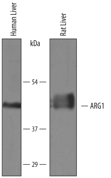
Detection of Human and Rat Arginase 1/ARG1 by Western Blot. Western blot shows lysates of human liver tissue and rat liver tissue. PVDF membrane was probed with 0.25 µg/mL of Sheep Anti-Human/Mouse/Rat Arginase 1/ARG1 Antigen Affinity-purified Polyclonal Antibody (Catalog # AF5868) followed by HRP-conjugated Anti-Sheep IgG Secondary Antibody (Catalog # HAF016). A specific band was detected for Arginase 1/ARG1 at approximately 41 kDa (as indicated). This experiment was conducted under reducing conditions and using Immunoblot Buffer Group 8.
 View Larger
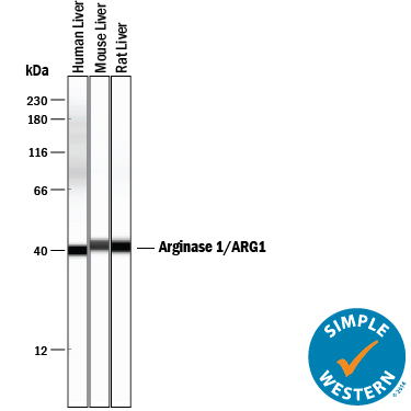
Detection of Human, Mouse, and Rat Arginase 1/ARG1 by Simple WesternTM. Simple Western lane view shows lysates of human liver tissue, mouse liver tissue, and rat liver tissue, loaded at 0.2 mg/mL. A specific band was detected for Arginase 1/ARG1 at approximately 41 kDa (as indicated) using 2.5 µg/mL of Sheep Anti-Human/Mouse/Rat Arginase 1/ARG1 Antigen Affinity-purified Polyclonal Antibody (Catalog # AF5868) followed by 1:50 dilution of HRP-conjugated Anti-Sheep IgG Secondary Antibody (Catalog # HAF016). This experiment was conducted under reducing conditions and using the 12-230 kDa separation system.
Reconstitution Calculator
Preparation and Storage
- 12 months from date of receipt, -20 to -70 °C as supplied.
- 1 month, 2 to 8 °C under sterile conditions after reconstitution.
- 6 months, -20 to -70 °C under sterile conditions after reconstitution.
Background: Arginase 1/ARG1
Arginase 1 (ARG1) is a 35‑40 kDa member of the arginase family of enzymes. It is expressed in multiple cell types, including erythrocytes, hepatocytes, neutrophils, smooth muscle and macrophages. ARG1 demonstrates two distinct functions: in the hepatocyte cytoplasm, it catalyzes the conversion of arginine to ornithine and urea, while in multiple cells, it degrades arginine, thus indirectly downregulating NO synthase (NOS) activity by depriving this enzyme of its substrate. Human AGR1 is 322 amino acids (aa) in length. Its enzyme region comprises aa 9‑309 and contains two Mn atoms. ARG1 is modestly active as a monomer, but highly active as a 105 kDa homotrimer. Trimerization is promoted by nitrosylation of Cys303, creating a regulatory feedback loop with NOS. There are two isoform variants, one that shows an eight aa insertion after Gln43, and another that shows a deletion of aa 204‑289. Full-length human ARG1 shares 87% aa identity with mouse and rat ARG1.
Product Datasheets
Citations for Human/Mouse/Rat Arginase 1/ARG1 Antibody
R&D Systems personnel manually curate a database that contains references using R&D Systems products. The data collected includes not only links to publications in PubMed, but also provides information about sample types, species, and experimental conditions.
12
Citations: Showing 1 - 10
Filter your results:
Filter by:
-
Leishmanicidal and immunomodulatory properties of Brazilian green propolis extract (EPP-AF�) and a gel formulation in a pre-clinical model
Authors: J Rebouças-S, NA Amorim, FH Jesus-Sant, JA de Lima, JB Lima, AA Berretta, VM Borges
Frontiers in Pharmacology, 2023-02-09;14(0):1013376.
Species: Mouse
Sample Types: Cell Lysates
Applications: Western Blot -
Essential roles of the cytokine oncostatin M in crosstalk between muscle fibers and immune cells in skeletal muscle after aerobic exercise
Authors: T Komori, Y Morikawa
The Journal of Biological Chemistry, 2022-11-09;0(0):102686.
Species: Mouse
Sample Types: Tissue Homogenates
Applications: Western Blot -
Cell-intrinsic Wnt4 ligand regulates mitochondrial oxidative phosphorylation in macrophages
Authors: M Tlili, H Acevedo, A Descoteaux, M Germain, KM Heinonen
The Journal of Biological Chemistry, 2022-06-25;0(0):102193.
Species: Mouse
Sample Types: Whole Cells
Applications: Flow Cytometry -
PD-L1 engagement on T cells promotes self-tolerance and suppression of neighboring macrophages and effector T cells in cancer
Authors: B Diskin, S Adam, MF Cassini, G Sanchez, M Liria, B Aykut, C Buttar, E Li, B Sundberg, RD Salas, R Chen, J Wang, M Kim, MS Farooq, S Nguy, C Fedele, KH Tang, T Chen, W Wang, M Hundeyin, JAK Rossi, E Kurz, MIU Haq, J Karlen, E Kruger, Z Sekendiz, D Wu, SAA Shadaloey, G Baptiste, G Werba, S Selvaraj, C Loomis, KK Wong, J Leinwand, G Miller
Nat. Immunol., 2020-03-09;21(4):442-454.
Species: Human, Mouse
Sample Types: Tissue Homogenates
Applications: Western Blot -
Extracellular vesicles secreted by hypoxia pre-challenged mesenchymal stem cells promote non-small cell lung cancer cell growth and mobility as well as macrophage M2 polarization via miR-21-5p delivery
Authors: W Ren, J Hou, C Yang, H Wang, S Wu, Y Wu, X Zhao, C Lu
J. Exp. Clin. Cancer Res., 2019-02-08;38(1):62.
Species: Xenograft
Sample Types: Cell Lysates
Applications: Western Blot -
The APMAP interactome reveals new modulators of APP processing and beta-amyloid production that are altered in Alzheimer's disease
Authors: H Gerber, S Mosser, B Boury-Jamo, M Stumpe, A Piersigill, C Goepfert, J Dengjel, U Albrecht, F Magara, PC Fraering
Acta Neuropathol Commun, 2019-01-31;7(1):13.
Species: Mouse
Sample Types: Tissue Homogenates
Applications: Western Blot -
Magnetic Field Changes Macrophage Phenotype
Authors: J Wosik, W Chen, K Qin, RM Ghobrial, JZ Kubiak, M Kloc
Biophys. J., 2018-04-24;114(8):2001-2013.
Species: Mouse
Sample Types: Cell Lysates
Applications: Western Blot -
Arginase1 Deficiency in Monocytes/Macrophages Upregulates Inducible Nitric Oxide Synthase To Promote Cutaneous Contact Hypersensitivity
Authors: J Suwanpradi, M Shih, L Pontius, B Yang, A Birukova, E Guttman-Ya, DL Corcoran, LG Que, RM Tighe, AS MacLeod
J. Immunol., 2017-07-26;0(0):.
Species: Mouse
Sample Types: Whole Cells
Applications: Flow Cytometry -
Assessment of Preclinical Liver and Skeletal Muscle Biomarkers Following Clofibrate Administration in Wistar Rats
Authors: P Maliver, M Festag, M Bennecke, F Christen, B Bánfai, B Lenz, M Winter
Toxicol Pathol, 2017-05-09;0(0):1926233177072.
Species: Rat
Sample Types: Serum
Applications: ELISA Development (Capture) -
IL-33 contributes to sepsis-induced long-term immunosuppression by expanding the regulatory T cell population
Authors: DC Nascimento, PH Melo, AR Piñeros, RG Ferreira, DF Colón, PB Donate, FV Castanheir, A Gozzi, PG Czaikoski, W Niedbala, MC Borges, DS Zamboni, FY Liew, FQ Cunha, JC Alves-Filh
Nat Commun, 2017-04-04;8(0):14919.
Species: Mouse
Sample Types: Cell Lysates
Applications: Western Blot -
Candida albicans Chitin Increases Arginase-1 Activity in Human Macrophages, with an Impact on Macrophage Antimicrobial Functions
Authors: J Wagener, DM MacCallum, GD Brown, NA Gow
MBio, 2017-01-24;8(1):.
Species: Human
Sample Types: Cell Lysates
Applications: Western Blot -
DAMP signaling is a key pathway inducing immune modulation after brain injury.
Authors: Liesz A, Dalpke A, Mracsko E, Antoine D, Roth S, Zhou W, Yang H, Na S, Akhisaroglu M, Fleming T, Eigenbrod T, Nawroth P, Tracey K, Veltkamp R
J Neurosci, 2015-01-14;35(2):583-98.
Species: Mouse
Sample Types: Cell Lysates
Applications: Western Blot
FAQs
No product specific FAQs exist for this product, however you may
View all Antibody FAQsReviews for Human/Mouse/Rat Arginase 1/ARG1 Antibody
Average Rating: 3.5 (Based on 2 Reviews)
Have you used Human/Mouse/Rat Arginase 1/ARG1 Antibody?
Submit a review and receive an Amazon gift card.
$25/€18/£15/$25CAN/¥75 Yuan/¥2500 Yen for a review with an image
$10/€7/£6/$10 CAD/¥70 Yuan/¥1110 Yen for a review without an image
Filter by:


