Human/Mouse/Rat beta-Catenin Antibody Summary
Ala2-Leu781
Accession # P35222
Applications
Please Note: Optimal dilutions should be determined by each laboratory for each application. General Protocols are available in the Technical Information section on our website.
Scientific Data
 View Larger
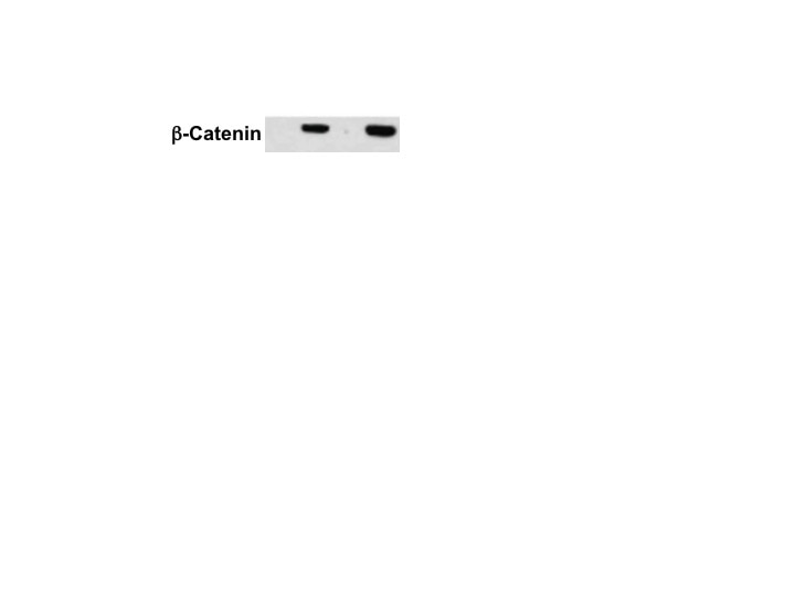
Detection of Human/Mouse/Rat beta ‑Catenin by Western Blot. Western blot shows lysates of HeLa human cervical epithelial carcinoma cell line, C6 rat glioma cell line, and NIH-3T3 mouse embryonic fibroblast cell line. PVDF membrane was probed with 1 µg/mL Goat Anti-Human/Mouse/Rat beta -Catenin Antigen Affinity-purified Polyclonal Antibody (Catalog # AF1329) followed by HRP-conjugated Anti-Goat IgG Secondary Antibody (Catalog # HAF109). For additional reference, recombinant human beta -catenin (1 ng) was included. A specific band for beta-Catenin was detected at approximately 95 kDa (as indicated). This experiment was conducted under reducing conditions and using Immunoblot Buffer Group 1.
 View Larger
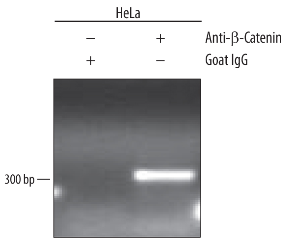
Detection of beta ‑Catenin-regul-ated Genes by Chromatin Immunoprecipitation. HeLa human cervical epithelial carcinoma cell line were fixed using formaldehyde, resuspended in lysis buffer, and sonicated to shear chromatin. beta -Catenin/ DNA complexes were immuno-precipitated using 5 µg Goat Anti-Human/Mouse/Rat beta -Catenin Antigen Affinity-purified Polyclonal Antibody (Catalog # AF1329) or control antibody (Catalog # AB-108-C) for 15 minutes in an ultrasonic bath, followed by Biotinylated Anti-Goat IgG Secondary Antibody (Catalog # BAF109). Immuno-complexes were captured using 50 µL of MagCellect Streptavidin Ferrofluid (Catalog # MAG999) and DNA was purified using chelating resin solution. TheSU(Z)12promoter was detected by standard PCR.
 View Larger
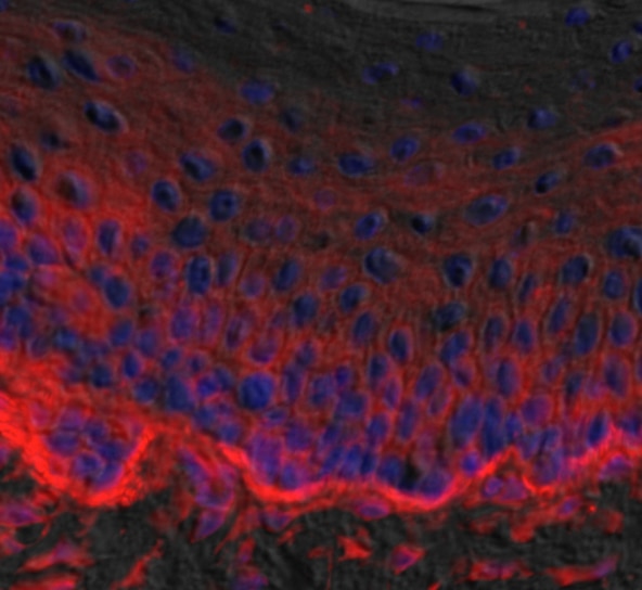
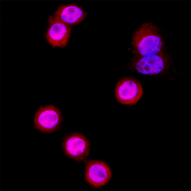
beta ‑Catenin in SW480 Human Cell Line. beta -Catenin was detected in immersion fixed SW480 human colorectal adenocarcinoma cell line using Goat Anti-Human/Mouse/Rat beta -Catenin Antigen Affinity-purified Polyclonal Antibody (Catalog # AF1329) at 15 µg/mL for 3 hours at room temperature. Cells were stained using the NorthernLights™ 557-conjugated Anti-Goat IgG Secondary Antibody (red; Catalog # NL001) and counterstained with DAPI (blue). Specific staining was localized to cytoplasm and nuclei. View our protocol for Fluorescent ICC Staining of Cells on Coverslips.
 View Larger
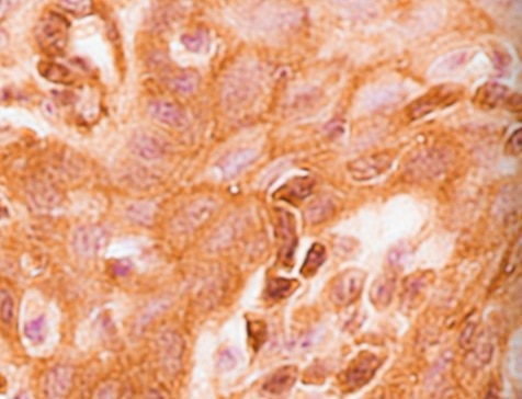
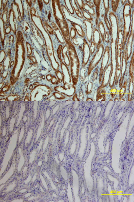
beta -Catenin in Human Kidney Cancer Tissue. beta -Catenin was detected in immersion fixed paraffin-embedded sections of human kidney cancer tissue using Goat Anti-Human/Mouse/Rat beta -Catenin Antigen Affinity-purified Polyclonal Antibody (Catalog # AF1329) at 15 µg/mL overnight at 4 °C. Tissue was stained using the Anti-Goat HRP-DAB Cell & Tissue Staining Kit (brown; Catalog # CTS008) and counterstained with hematoxylin (blue). Lower panel shows a lack of labeling if primary antibodies are omitted and tissue is stained only with secondary antibody followed by incubation with detection reagents. View our protocol for Chromogenic IHC Staining of Paraffin-embedded Tissue Sections.
 View Larger
beta ‑Catenin in Human Kidney Cancer Tissue. beta -Catenin was detected in immersion fixed paraffin-embedded sections of human kidney cancer tissue using 15 µg/mL Goat Anti-Human/Mouse/Rat beta -Catenin Antigen Affinity-purified Polyclonal Antibody (Catalog # AF1329) overnight at 4 °C. Tissue was stained with the Anti-Goat HRP-DAB Cell & Tissue Staining Kit (brown; Catalog # CTS008) and counterstained with hematoxylin (blue). Specific labeling was localized to epithelial cells in collecting tubules in the medulla. View our protocol for Chromogenic IHC Staining of Paraffin-embedded Tissue Sections.
 View Larger
Detection of beta ‑Catenin in HeLa Human Cell Line by Flow Cytometry. HeLa human cervical epithelial carcinoma cell line was stained with Goat Anti-Human/Mouse/Rat beta -Catenin Antigen Affinity-purified Polyclonal Antibody (Catalog # AF1329, filled histogram) or control antibody (Catalog # AB-108-C, open histogram), followed by Allophycocyanin-conjugated Anti-Goat IgG Secondary Antibody (Catalog # F0108). To facilitate intracellular staining, cells were fixed with paraformaldehyde and permeabilized with saponin.
 View Larger
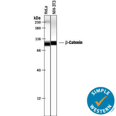
Detection of Human and Mouse beta ‑Catenin by Simple WesternTM. Simple Western lane view shows lysates of HeLa human cervical epithelial carcinoma cell line and NIH-3T3 mouse embryonic fibroblast cell line, loaded at 0.2 mg/mL. A specific band was detected for beta -Catenin at approximately 94-97 kDa (as indicated) using 50 µg/mL of Goat Anti-Human/Mouse/Rat beta -Catenin Antigen Affinity-purified Polyclonal Antibody (Catalog # AF1329) followed by 1:50 dilution of HRP-conjugated Anti-Goat IgG Secondary Antibody (Catalog # HAF109). This experiment was conducted under reducing conditions and using the 12-230 kDa separation system. Non-specific interaction with the 230 kDa Simple Western standard may be seen with this antibody.
Reconstitution Calculator
Preparation and Storage
- 12 months from date of receipt, -20 to -70 °C as supplied.
- 1 month, 2 to 8 °C under sterile conditions after reconstitution.
- 6 months, -20 to -70 °C under sterile conditions after reconstitution.
Background: beta-Catenin
beta -Catenin is a cadherin-associated protein that is involved in the regulation of cell adhesion. It also acts as a transcriptional co-activator in the nucleus and is involved the canonical Wnt signal transduction pathways.
Product Datasheets
Citations for Human/Mouse/Rat beta-Catenin Antibody
R&D Systems personnel manually curate a database that contains references using R&D Systems products. The data collected includes not only links to publications in PubMed, but also provides information about sample types, species, and experimental conditions.
20
Citations: Showing 1 - 10
Filter your results:
Filter by:
-
TCF/LEF regulation of the topologically associated domain ADI promotes mESCs to exit the pluripotent ground state
Authors: N Doumpas, S Söderholm, S Narula, S Moreira, BW Doble, C Cantù, K Basler
Cell Reports, 2021-09-14;36(11):109705.
Species: Mouse
Sample Types: Cell Lysates
Applications: Western Blot -
Shifting osteogenesis in vascular calcification
Authors: J Yao, X Wu, X Qiao, D Zhang, L Zhang, JA Ma, X Cai, KI Boström, Y Yao
JCI Insight, 2021-05-24;0(0):.
Species: Mouse
Sample Types: Cell Lysates, Tissue Homogenates
Applications: ChIP, ChIP-Seq, Western Blot -
LncRNA SNHG6 regulating Hedgehog signaling pathway and affecting the biological function of gallbladder carcinoma cells through targeting miR-26b-5p
Authors: XF Liu, K Wang, HC Du
Eur Rev Med Pharmacol Sci, 2020-07-01;24(14):7598-7611.
Species: Human
Sample Types: Cell Lysates
Applications: Western Blot -
The Actin-Family Protein Arp4 Is a Novel Suppressor for the Formation and Functions of Nuclear F-Actin
Authors: S Yamazaki, C Gerhold, K Yamamoto, Y Ueno, R Grosse, K Miyamoto, M Harata
Cells, 2020-03-19;9(3):.
Species: Mouse
Sample Types: Cell Culture Lysates
Applications: Immunoprecipitation -
Progranulin protects the mouse retina under hypoxic conditions via inhibition of the Toll?like receptor?4?NADPH oxidase 4 signaling pathway
Authors: ZP You, MJ Yu, YL Zhang, K Shi
Mol Med Rep, 2018-11-08;0(0):.
Species: Mouse
Sample Types: Tissue Homogenates
Applications: Western Blot -
Amnionless-mediated glycosylation is crucial for cell surface targeting of cubilin in renal and intestinal cells
Authors: T Udagawa, Y Harita, K Miura, J Mitsui, KL Ode, S Morishita, S Urae, S Kanda, Y Kajiho, H Tsurumi, HR Ueda, S Tsuji, A Saito, A Oka
Sci Rep, 2018-02-05;8(1):2351.
Species: Human
Sample Types: Whole Tissue
Applications: IHC-P -
WNT10A mutation causes ectodermal dysplasia by impairing progenitor cell proliferation and KLF4-mediated differentiation
Authors: M Xu, J Horrell, M Snitow, J Cui, H Gochnauer, CM Syrett, S Kallish, JT Seykora, F Liu, D Gaillard, JP Katz, KH Kaestner, B Levin, C Mansfield, JE Douglas, BJ Cowart, M Tordoff, F Liu, X Zhu, LA Barlow, AI Rubin, JA McGrath, EE Morrisey, EY Chu, SE Millar
Nat Commun, 2017-06-07;8(0):15397.
Species: Mouse
Sample Types: Whole Tissue
Applications: IHC-Fr -
Dchs1-Fat4 regulation of polarized cell behaviours during skeletal morphogenesis
Nat Commun, 2016-05-05;7(0):11469.
Species: Mouse
Sample Types: Whole Tissue
Applications: IHC -
ROS-induced endothelial stress contributes to pulmonary fibrosis through pericytes and Wnt signaling.
Authors: Andersson-Sjoland A, Karlsson J, Rydell-Tormanen K
Lab Invest, 2015-09-14;96(2):206-17.
Species: Mouse
Sample Types: Whole Tissue
Applications: IHC-P -
WNT1-induced Secreted Protein-1 (WISP1), a Novel Regulator of Bone Turnover and Wnt Signaling.
Authors: Maeda A, Ono M, Holmbeck K, Li L, Kilts T, Kram V, Noonan M, Yoshioka Y, McNerny E, Tantillo M, Kohn D, Lyons K, Robey P, Young M
J Biol Chem, 2015-04-11;290(22):14004-18.
Species: Mouse
Sample Types: Cell Lysates, Whole Tissue
Applications: IHC-P, Western Blot -
Integrin beta1 controls VE-cadherin localization and blood vessel stability.
Authors: Yamamoto H, Ehling M, Kato K, Kanai K, van Lessen M, Frye M, Zeuschner D, Nakayama M, Vestweber D, Adams R
Nat Commun, 2015-03-10;6(0):6429.
Species: Mouse
Sample Types: Tissue Homogenates
Applications: Immunoprecipitation, Western Blot -
Osteocytes mediate the anabolic actions of canonical Wnt/beta-catenin signaling in bone.
Authors: Tu X, Delgado-Calle J, Condon K, Maycas M, Zhang H, Carlesso N, Taketo M, Burr D, Plotkin L, Bellido T
Proc Natl Acad Sci U S A, 2015-01-20;112(5):E478-86.
Species: Mouse
Sample Types: Tissue Homogenates
Applications: Western Blot -
The Wnt/beta-catenin pathway attenuates experimental allergic airway disease.
Authors: Reuter S, Martin H, Beckert H, Bros M, Montermann E, Belz C, Heinz A, Ohngemach S, Sahin U, Stassen M, Buhl R, Eshkind L, Taube C
J Immunol, 2014-06-13;193(2):485-95.
Species: Mouse
Sample Types: Whole Tissue
Applications: IHC-P -
High glucose induces podocyte injury via enhanced (pro)renin receptor-Wnt-beta-catenin-snail signaling pathway.
Authors: Li C, Siragy H
PLoS ONE, 2014-02-12;9(2):e89233.
Species: Mouse
Sample Types: Cell Lysates
Applications: Western Blot -
Shaping organs by a wingless-int/Notch/nonmuscle myosin module which orients feather bud elongation.
Authors: Li A, Chen M, Jiang T, Wu P, Nie Q, Widelitz R, Chuong C
Proc Natl Acad Sci U S A, 2013-04-01;110(16):E1452-61.
Species: Chicken
Sample Types: Whole Tissue
Applications: Bioassay -
Canonical Wnt signaling negatively regulates platelet function.
Authors: Steele BM, Harper MT, Macaulay IC, Morrell CN, Perez-Tamayo A, Foy M, Habas R, Poole AW, Fitzgerald DJ, Maguire PB
Proc. Natl. Acad. Sci. U.S.A., 2009-11-09;106(47):19836-41.
Species: Human
Sample Types: Cell Lysates
Applications: Western Blot -
Blockade of Wnt signaling inhibits angiogenesis and tumor growth in hepatocellular carcinoma.
Authors: Hu J, Dong A, Fernandez-Ruiz V, Shan J, Kawa M, Martinez-Anso E, Prieto J, Qian C
Cancer Res., 2009-08-18;69(17):6951-9.
Species: Human
Sample Types: Tissue Homogenates
Applications: Western Blot -
Mutational analysis of the FXNPXY motif within LDL receptor-related protein 1 (LRP1) reveals the functional importance of the tyrosine residues in cell growth regulation and signal transduction.
Authors: Zhang H, Lee JM, Wang Y, Dong L, Ko KW, Pelletier L, Yao Z
Biochem. J., 2008-01-01;409(1):53-64.
Species: Hamster
Sample Types: Cell Lysates
Applications: Western Blot -
Dickkopf-1 is a master regulator of joint remodeling.
Authors: Diarra D, Stolina M, Polzer K, Zwerina J, Ominsky MS, Dwyer D, Korb A, Smolen J, Hoffmann M, Scheinecker C, van der Heide D, Landewe R, Lacey D, Richards WG, Schett G
Nat. Med., 2007-01-21;13(2):156-63.
Species: Human, Mouse
Sample Types: Tissue Homogenates
Applications: Western Blot -
Requirements for proximal tubule epithelial cell detachment in response to ischemia: role of oxidative stress.
Authors: Saenz-Morales D, Escribese MM, Stamatakis K, Garcia-Martos M, Alegre L, Conde E, Perez-Sala D, Mampaso F, Garcia-Bermejo ML
Exp. Cell Res., 2006-09-12;312(19):3711-27.
Species: Rat
Sample Types: Whole Cells
Applications: ICC
FAQs
No product specific FAQs exist for this product, however you may
View all Antibody FAQsReviews for Human/Mouse/Rat beta-Catenin Antibody
Average Rating: 4.5 (Based on 4 Reviews)
Have you used Human/Mouse/Rat beta-Catenin Antibody?
Submit a review and receive an Amazon gift card.
$25/€18/£15/$25CAN/¥75 Yuan/¥2500 Yen for a review with an image
$10/€7/£6/$10 CAD/¥70 Yuan/¥1110 Yen for a review without an image
Filter by:
