Human/Mouse/Rat Calreticulin Antibody Summary
Glu18-Leu417
Accession # P27797
Applications
Please Note: Optimal dilutions should be determined by each laboratory for each application. General Protocols are available in the Technical Information section on our website.
Scientific Data
 View Larger
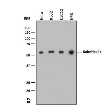
Detection of Human, Mouse, and Rat Calreticulin by Western Blot. Western blot shows lysates of HeLa human cervical epithelial carcinoma cell line, K562 human chronic myelogenous leukemia cell line, C2C12 mouse myoblast cell line, and NRK rat normal kidney cell line. PVDF membrane was probed with 0.5 µg/mL of Mouse Anti-Human Calreticulin Monoclonal Antibody (Catalog # MAB38981) followed by HRP-conjugated Anti-Mouse IgG Secondary Antibody (Catalog # HAF018). A specific band was detected for Calreticulin at approximately 55 kDa (as indicated). This experiment was conducted under reducing conditions and using Immunoblot Buffer Group 1.
 View Larger
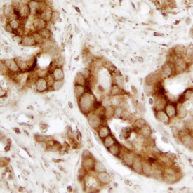
Calreticulin in Human Prostate. Calreticulin was detected in formalin fixed paraffin-embedded sections of human prostate using Mouse Anti-Human Calreticulin Monoclonal Antibody (Catalog # MAB38981) at 15 µg/mL overnight at 4 °C. Before incubation with the primary antibody, tissue was subjected to heat-induced epitope retrieval using Antigen Retrieval Reagent-Basic (Catalog # CTS013). Tissue was stained using the Anti-Mouse HRP-DAB Cell & Tissue Staining Kit (brown; Catalog # CTS002) and counterstained with hematoxylin (blue). Specific staining was localized to plasma membrane and cytoplasm. View our protocol for Chromogenic IHC Staining of Paraffin-embedded Tissue Sections.
 View Larger
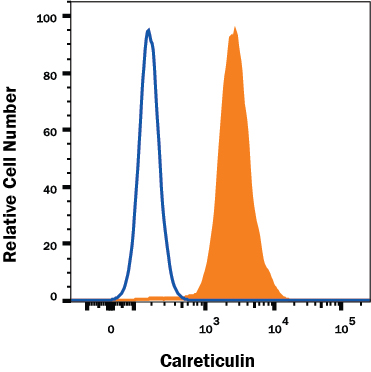
Detection of Calreticulin in HeLa Human Cell Line by Flow Cytometry. HeLa human cervical epithelial carcinoma cell line was stained with Mouse Anti-Human Calreticulin Monoclonal Antibody (Catalog # MAB38981, filled histogram) or isotype control antibody (Catalog # MAB0041, open histogram), followed by Allophycocyanin-conjugated Anti-Mouse IgG Secondary Antibody (Catalog # F0101B). To facilitate intracellular staining, cells were fixed with Flow Cytometry Fixation Buffer (Catalog # FC004) and permeabilized with Flow Cytometry Permeabilization/Wash Buffer I (Catalog # FC005). View our protocol for Staining Intracellular Molecules.
 View Larger
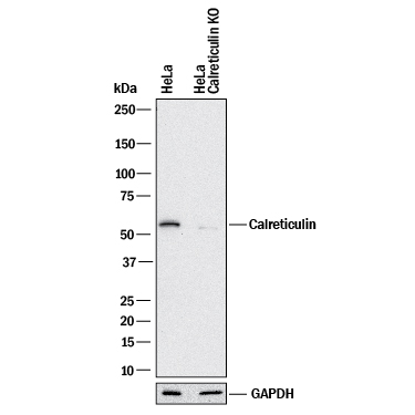
Western Blot Shows Human Calreticulin Specificity by Using Knockout Cell Line. Western blot shows lysates of HeLa human cervical epithelial carcinoma parental cell line and Calreticulin knockout HeLa cell line (KO). PVDF membrane was probed with 0.5 µg/mL of Mouse Anti-Human/Mouse/Rat Calreticulin Monoclonal Antibody (Catalog # MAB38981) followed by HRP-conjugated Anti-Mouse IgG Secondary Antibody (Catalog # HAF018). A specific band was detected for Calreticulin at approximately 55 kDa (as indicated) in the parental HeLa cell line, but is not detectable in knockout HeLa cell line. GAPDH (Catalog # MAB5718) is shown as a loading control. This experiment was conducted under reducing conditions and using Immunoblot Buffer Group 1.
Reconstitution Calculator
Preparation and Storage
- 12 months from date of receipt, -20 to -70 °C as supplied.
- 1 month, 2 to 8 °C under sterile conditions after reconstitution.
- 6 months, -20 to -70 °C under sterile conditions after reconstitution.
Background: Calreticulin
Human Calreticulin is a 55‑60 kDa, 400 amino acid, variably glycosylated intra- and extracellular Ca++-binding lectin that is ubiquitously expressed. It consists of three domains: a 180 aa N-terminal globular region, a 111 aa P-, or proline rich domain, and a 109 aa C-terminus. The 180 aa N-terminus (aa 18‑197) is termed Vasostatin. It is unclear if it is ever generated naturally via proteolytic processing. Vasostatin domain has many functions. It binds to RNA (aa 18‑27), has autocatalytic phosphorylase activity (aa 77‑197), binds to a KxFFKR motif on steroid hormone receptors, and serves as a lectin-type chaperone for ER-localized molecules. It also shows antiangiogenic activity, presumably by binding to laminin carbohydrates and blocking endothelial cell adhesion and proliferation. Human Calreticulin is 94% aa identical to mouse and rat Calreticulin.
Product Datasheets
Citations for Human/Mouse/Rat Calreticulin Antibody
R&D Systems personnel manually curate a database that contains references using R&D Systems products. The data collected includes not only links to publications in PubMed, but also provides information about sample types, species, and experimental conditions.
2
Citations: Showing 1 - 2
Filter your results:
Filter by:
-
The NK�cell receptor NKp46 recognizes ecto-calreticulin on ER-stressed cells
Authors: S Sen Santar, DJ Lee, Â Crespo, JJ Hu, C Walker, X Ma, Y Zhang, S Chowdhury, KF Meza-Sosa, M Lewandrows, H Zhang, M Rowe, A McClelland, H Wu, C Junqueira, J Lieberman
Nature, 2023-04-05;616(7956):348-356.
Species: Human
Sample Types: Whole Cells
Applications: ICC -
Reversing insufficient photothermal therapy-induced tumor relapse and metastasis by regulating cancer-associated fibroblasts
Authors: X Li, T Yong, Z Wei, N Bie, X Zhang, G Zhan, J Li, J Qin, J Yu, B Zhang, L Gan, X Yang
Nature Communications, 2022-05-19;13(1):2794.
Species: Human, Mouse
Sample Types: Whole Cells
Applications: Flow Cytometry
FAQs
No product specific FAQs exist for this product, however you may
View all Antibody FAQsReviews for Human/Mouse/Rat Calreticulin Antibody
There are currently no reviews for this product. Be the first to review Human/Mouse/Rat Calreticulin Antibody and earn rewards!
Have you used Human/Mouse/Rat Calreticulin Antibody?
Submit a review and receive an Amazon gift card.
$25/€18/£15/$25CAN/¥75 Yuan/¥2500 Yen for a review with an image
$10/€7/£6/$10 CAD/¥70 Yuan/¥1110 Yen for a review without an image

