Human/Mouse/Rat FAK Antibody Summary
Asp213-Thr412
Accession # Q05397
Applications
Please Note: Optimal dilutions should be determined by each laboratory for each application. General Protocols are available in the Technical Information section on our website.
Scientific Data
 View Larger
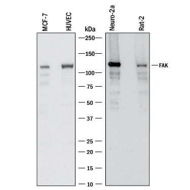
Detection of Human, Mouse, and Rat FAK by Western Blot. Western blot shows lysates of MCF‑7 human breast cancer cell line, HUVEC human umbilical vein endothelial cells, Neuro‑2A mouse neuroblastoma cell line, and Rat‑2 rat embryonic fibroblast cell line. PVDF membrane was probed with 1 µg/mL of Sheep Anti-Human/Mouse/Rat FAK Antigen Affinity-purified Polyclonal Antibody (Catalog # AF4467) followed by HRP-conjugated Anti-Sheep IgG Secondary Antibody (HAF016). A specific band was detected for FAK at approximately 125 kDa (as indicated). This experiment was conducted under reducing conditions and using Western Blot Buffer Group 1.
 View Larger
FAK in Human Brain. FAK was detected in immersion fixed paraffin-embedded sections of human brain (hippocampus) using 3 µg/mL Human/Mouse/Rat FAK Antigen Affinity-purified Polyclonal Antibody (Catalog # AF4467) overnight at 4 °C. Tissue was stained with the Anti-Sheep HRP-DAB Cell & Tissue Staining Kit (brown; Catalog # CTS019) and counterstained with hematoxylin (blue). View our protocol for Chromogenic IHC Staining of Paraffin-embedded Tissue Sections.
 View Larger
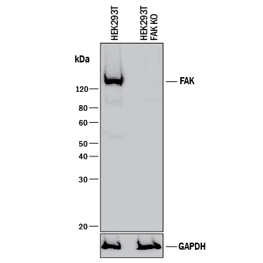
Western Blot Shows Human FAK Specificity by Using Knockout Cell Line. Western blot shows lysates of HEK293T human embryonic kidney parental cell line and FAK knockout HEK293T cell line (KO). PVDF membrane was probed with 1 µg/mL of Sheep Anti-Human/Mouse/Rat FAK Antigen Affinity-purified Polyclonal Antibody (Catalog # AF4467) followed by HRP-conjugated Anti-Sheep IgG Secondary Antibody (Catalog # HAF016). A specific band was detected for FAK at approximately 135 kDa (as indicated) in the parental HEK293T cell line, but is not detectable in knockout HEK293T cell line. GAPDH (Catalog # AF5718) is shown as a loading control. This experiment was conducted under reducing conditions and using Immunoblot Buffer Group 1.
Reconstitution Calculator
Preparation and Storage
- 12 months from date of receipt, -20 to -70 °C as supplied.
- 1 month, 2 to 8 °C under sterile conditions after reconstitution.
- 6 months, -20 to -70 °C under sterile conditions after reconstitution.
Background: FAK
Focal adhesion kinase 1 (FAK) is a ubiquitously expressed non-receptor protein tyrosine kinase that is concentrated in the focal adhesions that form between cells growing in the presence of extracellular matrix constituents. This cellular localization is directed by a "Focal Adhesion Targeting" (FAT) sequence, a 125 amino acid sequence at the C-terminus. FAK plays an important role in migration, cell spreading, differentiation, cytoskeleton protein phosphorylation, apoptosis and acceleration of the G1 to S phase transition of the cell cycle. It associates with several different signaling proteins such as Src-family PTKs, p130Cas, Shc, Grb2, PI 3-kinase, and paxillin. This enables FAK to function within a network of integrin-stimulated signaling pathways leading to the activation of targets such as the ERK and JNK/mitogen-activated protein kinase pathways. FAK is also linked to oncogenes at biochemical and functional levels. Increased expression and/or activity of FAK in various tumors has been correlated with enhanced migration and invasiveness of human tumor cells in addition to promoting increased cell proliferation.
Product Datasheets
Citations for Human/Mouse/Rat FAK Antibody
R&D Systems personnel manually curate a database that contains references using R&D Systems products. The data collected includes not only links to publications in PubMed, but also provides information about sample types, species, and experimental conditions.
2
Citations: Showing 1 - 2
Filter your results:
Filter by:
-
Focal adhesion kinase coordinates costamere-related JNK signaling with muscle fiber transformation after Achilles tenotomy and tendon reconstruction
Authors: C Ferrié, S Kasper, F Wanivenhau, M Flück
Exp. Mol. Pathol., 2019-03-15;0(0):.
Species: Rat
Sample Types: Tissue Homogenates
Applications: ELISA Development (Capture) -
Knee Extensors Muscle Plasticity Over a 5-Years Rehabilitation Process After Open Knee Surgery
Authors: M Flück, C Viecelli, AM Bapst, S Kasper, P Valdivieso, MV Franchi, S Ruoss, JM Lüthi, M Bühler, H Claassen, H Hoppeler, C Gerber
Front Physiol, 2018-09-25;9(0):1343.
Species: Human
Sample Types: Tissue Homogenates
Applications: Immunoprecipitation
FAQs
No product specific FAQs exist for this product, however you may
View all Antibody FAQsReviews for Human/Mouse/Rat FAK Antibody
There are currently no reviews for this product. Be the first to review Human/Mouse/Rat FAK Antibody and earn rewards!
Have you used Human/Mouse/Rat FAK Antibody?
Submit a review and receive an Amazon gift card.
$25/€18/£15/$25CAN/¥75 Yuan/¥2500 Yen for a review with an image
$10/€7/£6/$10 CAD/¥70 Yuan/¥1110 Yen for a review without an image


