Human/Mouse/Rat Galectin-3 Antibody Summary
Ala2-Ile264
Accession # NP_034835
Applications
Please Note: Optimal dilutions should be determined by each laboratory for each application. General Protocols are available in the Technical Information section on our website.
Scientific Data
 View Larger
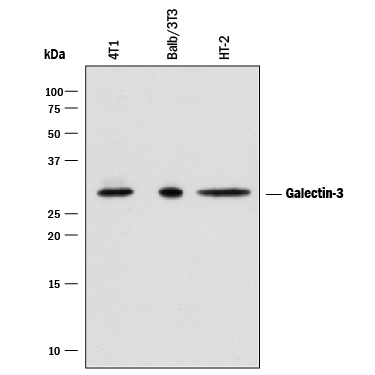
Detection of Mouse Galectin‑3 by Western Blot. Western blot shows lysates of 4T1 mouse breast cancer cell line, Balb/3T3 mouse embryonic fibroblast cell line and HT-2 mouse T cell line. PVDF membrane was probed with 0.05 µg/mL of Goat Anti-Mouse Galectin-3 Antigen Affinity-purified Polyclonal Antibody (Catalog # AF1197) followed by HRP-conjugated Anti-Goat IgG Secondary Antibody (Catalog # HAF017). A specific band was detected for Galectin-3 at approximately 28 kDa (as indicated). This experiment was conducted under reducing conditions and using Immunoblot Buffer Group 1.
 View Larger
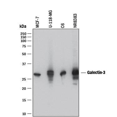
Detection of Human and Rat Galectin‑3 by Western Blot. Western blot shows lysates of MCF-7 human breast cancer cell line, U-118-MG human glioblastoma/astrocytoma cell line, C6 rat glioma cell line, and NR8383 rat alveolar macrophage cell line. PVDF membrane was probed with 0.2 µg/mL of Goat Anti-Mouse Galectin-3 Antigen Affinity-purified Polyclonal Antibody (Catalog # AF1197) followed by HRP-conjugated Anti-Goat IgG Secondary Antibody (Catalog # HAF017). A specific band was detected for Galectin-3 at approximately 28 kDa (as indicated). This experiment was conducted under reducing conditions and using Immunoblot Buffer Group 1.
 View Larger
Galectin‑3 in Mouse Embryo. Galectin-3 was detected in immersion fixed frozen sections of mouse embryo (15 d.p.c., cross-section through the eye) using Goat Anti-Human Galectin-3 Antigen Affinity-purified Polyclonal Antibody (Catalog # AF1197 ) at 15 µg/mL overnight at 4 °C. Tissue was stained using the Anti-Goat HRP-DAB Cell & Tissue Staining Kit (brown; Catalog # CTS008) and counterstained with hematoxylin (blue). View our protocol for Chromogenic IHC Staining of Frozen Tissue Sections.
 View Larger
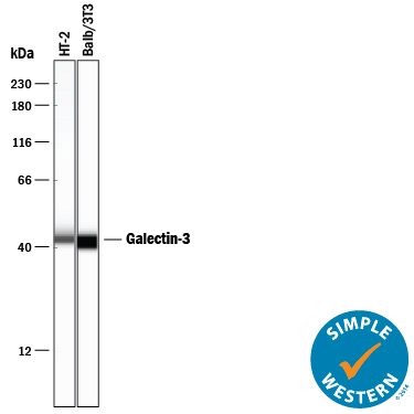
Detection of Mouse Galectin‑3 by Simple WesternTM. Simple Western lane view shows lysates of HT-2 mouse T cell line and Balb/3T3 mouse embryonic fibroblast cell line, loaded at 0.2 mg/mL. A specific band was detected for Galectin-3 at approximately 44 kDa (as indicated) using 10 µg/mL of Goat Anti-Mouse Galectin-3 Antigen Affinity-purified Polyclonal Antibody (Catalog # AF1197) followed by 1:50 dilution of HRP-conjugated Anti-Goat IgG Secondary Antibody (Catalog # HAF109). This experiment was conducted under reducing conditions and using the 12-230 kDa separation system.
Reconstitution Calculator
Preparation and Storage
- 12 months from date of receipt, -20 to -70 °C as supplied.
- 1 month, 2 to 8 °C under sterile conditions after reconstitution.
- 6 months, -20 to -70 °C under sterile conditions after reconstitution.
Background: Galectin-3
The galectins constitute a large family of carbohydrate-binding proteins with specificity for N-acetyl-lactosamine-containing glycoproteins. At least 14 mammalian galectins, which share structural similarities in their carbohydrate recognition domains (CRD), have been identified. The galectins have been classified into the prototype galectins (-1, -2, -5, -7, -10, -11, -13, -14), which contain one CRD and exist either as a monomer or a noncovalent homodimer; the chimera galectins (Galectin-3) containing one CRD linked to a nonlectin domain; and the tandem-repeat galectins (-4, -6, -8, -9, -12) consisting of two CRDs joined by a linker peptide. Galectins lack a classical signal peptide and can be localized to the cytosolic compartments where they have intracellular functions. However, via one or more as yet unidentified non-classical secretory pathways, galectins can also be secreted to function extracellularly. Individual members of the galectin family have different tissue distribution profiles and exhibit subtle differences in their carbohydrate-binding specificities. Each family member may preferentially bind to a unique subset of cell-surface glycoproteins (1-4).
Galectin-3, also known as Mac-2, L29, CBP35, and epsilon BP, is a chimera galectin that has a tendency to dimerize. Besides the soluble protein, alternatively spliced forms of chicken Galectin-3 containing a transmembrane-spanning domain and a leucine zipper motif have been reported. Galectin-3 is expressed in tumor cells, macrophages, activated T cells, osteoclasts, epithelial cells, and fibroblasts. It binds various matrix glycoproteins including laminin, fibronectin, LAMPS, 90K/Mac-2BP, MP20, and CEA. Galectin-3 promotes cell growth and proliferation for many cell types. Galectin-3 acts intracellularly to prevent apoptosis. Depending on the cell types, Galectin-3 exhibits pro- or anti-adhesive properties. Galectin-3 has proinflammatory activities in vitro and in vivo. It induces pro-inflammatory and inhibits Th2 type cytokine production. Galectin-3 chemoattracts monocytes and macrophages. It activates and degranulates basophils and mast cells. Elevated circulating levels of Galectin-3 has been show to correlate with the malignant potential of several types of cancer, suggesting that Galectin-3 is also involved in tumor growth and metastasis. Human and mouse Galectin-3 shares approximately 80% amino acid sequence similarity (1-5).
- Rabinovich, A. et al. (2002) Trends in Immunol. 23:313.
- Rabinovich, A. et al. (2002) J. Leukocyte Biology 71:741.
- Hughes, R.C. (2001) Biochimie 83:667.
- R&D Systems Cytokine Bulletin; Summer 2002.
- Gorski, J.P. et al. (2002) J. Biol. Chem. 277:18840.
Product Datasheets
Citations for Human/Mouse/Rat Galectin-3 Antibody
R&D Systems personnel manually curate a database that contains references using R&D Systems products. The data collected includes not only links to publications in PubMed, but also provides information about sample types, species, and experimental conditions.
22
Citations: Showing 1 - 10
Filter your results:
Filter by:
-
Equilibrative nucleoside transporter 3 supports microglial functions and protects against the progression of Huntington's disease in the mouse model
Authors: Lu, YS;Hung, WC;Hsieh, YT;Tsai, PY;Tsai, TH;Fan, HH;Chang, YG;Cheng, HK;Huang, SY;Lin, HC;Lee, YH;Shen, TH;Hung, BY;Tsai, JW;Dzhagalov, I;Cheng, IH;Lin, CJ;Chern, Y;Hsu, CL;
Brain, behavior, and immunity
Species: Mouse
Sample Types: Whole Tissue
Applications: Immunohistochemistry -
Mutation of the galectin-3 glycan-binding domain (Lgals3-R200S) enhances cortical bone expansion in male mice and trabecular bone mass in female mice
Authors: KA Maupin, CR Diegel, PD Stevens, D Dick, VAI Vivari, BO Williams
FEBS Open Bio, 2022-09-14;0(0):.
Species: Transgenic Mouse
Sample Types: Cell Lysates, Whole Cells
Applications: ICC, Western Blot -
Regulating microglial miR-155 transcriptional phenotype alleviates Alzheimer's-induced retinal vasculopathy by limiting Clec7a/Galectin-3+ neurodegenerative microglia
Authors: H Shi, Z Yin, Y Koronyo, DT Fuchs, J Sheyn, MR Davis, JW Wilson, MA Margeta, KM Pitts, S Herron, S Ikezu, T Ikezu, SL Graham, VK Gupta, KL Black, M Mirzaei, O Butovsky, M Koronyo-Ha
Acta neuropathologica communications, 2022-09-08;10(1):136.
Species: Mouse
Sample Types: Whole Tissue
Applications: IHC -
Crosstalk between dihydroceramides produced by Porphyromonas gingivalis and host lysosomal cathepsin B in the promotion of osteoclastogenesis
Authors: C Duarte, C Yamada, C Garcia, J Akkaoui, A Ho, F Nichols, A Movila
Journal of Cellular and Molecular Medicine, 2022-04-16;0(0):.
Species: Mouse
Sample Types: Cell Lysates, Whole Cells
Applications: IFC, Western Blot -
Astrocytic phagocytosis contributes to demyelination after focal cortical ischemia in mice
Authors: T Wan, W Zhu, Y Zhao, X Zhang, R Ye, M Zuo, P Xu, Z Huang, C Zhang, Y Xie, X Liu
Nature Communications, 2022-03-03;13(1):1134.
Species: Mouse
Sample Types: Whole Cells
Applications: ICC -
Gal3 Plays a Deleterious Role in a Mouse Model of Endotoxemia
Authors: JC Fernández-, AM Espinosa-O, I García-Dom, I Rosado-Sán, YM Pacheco, R Moyano, JG Monterde, JL Venero, RM de Pablos
International Journal of Molecular Sciences, 2022-01-21;23(3):.
Species: Mouse
Sample Types: Whole Tissue
Applications: IHC -
Age-related changes in brain phospholipids and bioactive lipids in the APP knock-in mouse model of Alzheimer's disease
Authors: C Emre, KV Do, B Jun, E Hjorth, SG Alcalde, MI Kautzmann, WC Gordon, P Nilsson, NG Bazan, M Schultzber
Acta neuropathologica communications, 2021-06-29;9(1):116.
Species: Mouse
Sample Types: Tissue Homogenates
Applications: Western Blot -
Diurnal Photoreceptor Outer Segment Renewal in Mice Is Independent of Galectin-3
Authors: NJ Esposito, F Mazzoni, JA Vargas, SC Finnemann
Investigative Ophthalmology & Visual Science, 2021-02-01;62(2):7.
Species: Mouse
Sample Types: Whole Tissue
Applications: IHC -
Network analysis of the progranulin-deficient mouse brain proteome reveals pathogenic mechanisms shared in human frontotemporal dementia caused by GRN mutations
Authors: M Huang, E Modeste, E Dammer, P Merino, G Taylor, DM Duong, Q Deng, CJ Holler, M Gearing, D Dickson, NT Seyfried, T Kukar
Acta Neuropathol Commun, 2020-10-07;8(1):163.
Species: Mouse
Sample Types: Protein, Whole Tissue
Applications: IHC-P, Western Blot -
Serum amyloid A-containing HDL binds adipocyte-derived versican and macrophage-derived biglycan, reducing its anti-inflammatory properties
Authors: CY Han, I Kang, M Omer, S Wang, T Wietecha, TN Wight, A Chait
JCI Insight, 2020-09-24;0(0):.
Species: Human, Mouse
Sample Types: Whole Tissue
Applications: IHC -
Plasma APE1/Ref-1 Correlates with Atherosclerotic Inflammation in ApoE-/- Mice
Authors: YR Lee, HK Joo, EO Lee, MS Park, HS Cho, S Kim, H Jin, JO Jeong, CS Kim, BH Jeon
Biomedicines, 2020-09-21;8(9):.
Species: Mouse
Sample Types: Tissue Homogenates, Whole Cells, Whole Tissue
Applications: ICC, IHC, Western Blot -
Adipocyte-Derived Versican and Macrophage-Derived Biglycan Control Adipose Tissue Inflammation in Obesity
Authors: CY Han, I Kang, IA Harten, JA Gebe, CK Chan, M Omer, KM Alonge, LJ den Hartig, D Gomes Kjer, L Goodspeed, S Subramania, S Wang, F Kim, DE Birk, TN Wight, A Chait
Cell Rep, 2020-06-30;31(13):107818.
Species: Mouse
Sample Types: Whole Tissue
Applications: IHC -
Voluntary running does not reduce neuroinflammation or improve non-cognitive behavior in the 5xFAD mouse model of Alzheimer's disease
Authors: M Svensson, E Andersson, O Manouchehr, Y Yang, T Deierborg
Sci Rep, 2020-01-28;10(1):1346.
Species: Mouse
Sample Types: Cell Lysates, Whole Tissue
Applications: IHC, Western Blot -
Bush-like integrin filament networks associated with hyaloid vasculature in murine neonate eyes
Authors: T Iwanaga, J Nio-Kobaya, H Takahashi-
Biomed. Res., 2019-01-01;40(2):79-85.
Species: Mouse
Sample Types: Whole Tissue
Applications: IHC -
Peripheral Inflammation Enhances Microglia Response and Nigral Dopaminergic Cell Death in an in vivo MPTP Model of Parkinson's Disease
Authors: I García-Dom, K Veselá, J García-Rev, A Carrillo-J, MA Roca-Cebal, M Santiago, RM de Pablos, JL Venero
Front Cell Neurosci, 2018-11-06;12(0):398.
Species: Mouse
Sample Types: Whole Tissue
Applications: IHC-Fr -
Acute Complement Inhibition Potentiates Neurorehabilitation and Enhances tPA-Mediated Neuroprotection
Authors: A Alawieh, M Andersen, DL Adkins, S Tomlinson
J. Neurosci., 2018-06-19;38(29):6527-6545.
Species: Mouse
Sample Types: Whole Tissue
Applications: IHC -
Histochemical characteristics of regressing vessels in the hyaloid vascular system of neonatal mice: Novel implication for vascular atrophy
Authors: A Kishimoto, S Kimura, J Nio-Kobaya, H Takahashi-, AM Park, T Iwanaga
Exp. Eye Res., 2018-03-27;172(0):1-9.
Species: Mouse
Sample Types: Whole Tissue
Applications: IHC -
Identifying the role of complement in triggering neuroinflammation after traumatic brain injury
Authors: A Alawieh, EF Langley, S Weber, D Adkins, S Tomlinson
J. Neurosci., 2018-02-06;0(0):.
Species: Mouse
Sample Types: Whole Tissue
Applications: IHC -
Kr�ppel-like factor 3 (KLF3/BKLF) is required for widespread repression of the inflammatory modulator galectin-3 (Lgals3)
J Biol Chem, 2016-05-24;0(0):.
Species: Mouse
Sample Types: Cell Culture Supernates, Whole Tissue
Applications: IHC-P, Western Blot -
Galectin-1 and galectin-8 have redundant roles in promoting plasma cell formation.
Authors: Tsai CM, Guan CH, Hsieh HW, Hsu TL, Tu Z, Wu KJ, Lin CH, Lin KI
J. Immunol., 2011-07-13;187(4):1643-52.
Species: Mouse
Sample Types: Cell Lysates
Applications: Western Blot -
Characterization of RAGE, HMGB1, and S100beta in inflammation-induced preterm birth and fetal tissue injury.
Authors: Buhimschi CS, Baumbusch MA, Dulay AT, Oliver EA, Lee S, Zhao G, Bhandari V, Ehrenkranz RA, Weiner CP, Madri JA, Buhimschi IA
Am. J. Pathol., 2009-08-13;175(3):958-75.
Species: Human
Sample Types: Whole Tissue
Applications: IHC-P -
Galectin-3 is an amplifier of inflammation in atherosclerotic plaque progression through macrophage activation and monocyte chemoattraction.
Authors: Papaspyridonos M, McNeill E, de Bono JP, Smith A, Burnand KG, Channon KM, Greaves DR
Arterioscler. Thromb. Vasc. Biol., 2007-12-20;28(3):433-40.
Species: Mouse
Sample Types: Tissue Homogenates, Whole Tissue
Applications: IHC-P, Western Blot
FAQs
No product specific FAQs exist for this product, however you may
View all Antibody FAQsReviews for Human/Mouse/Rat Galectin-3 Antibody
There are currently no reviews for this product. Be the first to review Human/Mouse/Rat Galectin-3 Antibody and earn rewards!
Have you used Human/Mouse/Rat Galectin-3 Antibody?
Submit a review and receive an Amazon gift card.
$25/€18/£15/$25CAN/¥75 Yuan/¥2500 Yen for a review with an image
$10/€7/£6/$10 CAD/¥70 Yuan/¥1110 Yen for a review without an image

