Human/Mouse/Rat N-Cadherin Antibody Summary
(rh) E-Cadherin, and rhR-Cadherin is observed.
Asp160-Ala724
Accession # P19022
Applications
This antibody functions as an ELISA detection antibody to detect human N-Cadherin when paired with Mouse Anti-Human N‑Cadherin Monoclonal Antibody (Catalog # MAB13883).
This product is intended for assay development on various assay platforms requiring antibody pairs. We recommend the Human N-Cadherin DuoSet ELISA Kit (Catalog # DY1388-05) for convenient development of a sandwich ELISA.
Please Note: Optimal dilutions should be determined by each laboratory for each application. General Protocols are available in the Technical Information section on our website.
Scientific Data
 View Larger
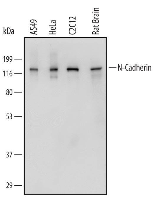
Detection of Human, Mouse, and Rat N‑Cadherin by Western Blot. Western blot shows lysates of A549 human lung carcinoma cell line, HeLa human cervical epithelial carcinoma cell line, C2C12 mouse myoblast cell line, and rat brain tissue. PVDF Membrane was probed with 0.5 µg/mL of Sheep Anti-Human/Mouse/Rat N-Cadherin Antigen Affinity-purified Polyclonal Antibody (Catalog # AF6426) followed by HRP-conjugated Anti-Sheep IgG Secondary Antibody (Catalog # HAF016). A specific band was detected for N-Cadherin at approximately 130 kDa (as indicated). This experiment was conducted under reducing conditions and using Immunoblot Buffer Group 1.
 View Larger
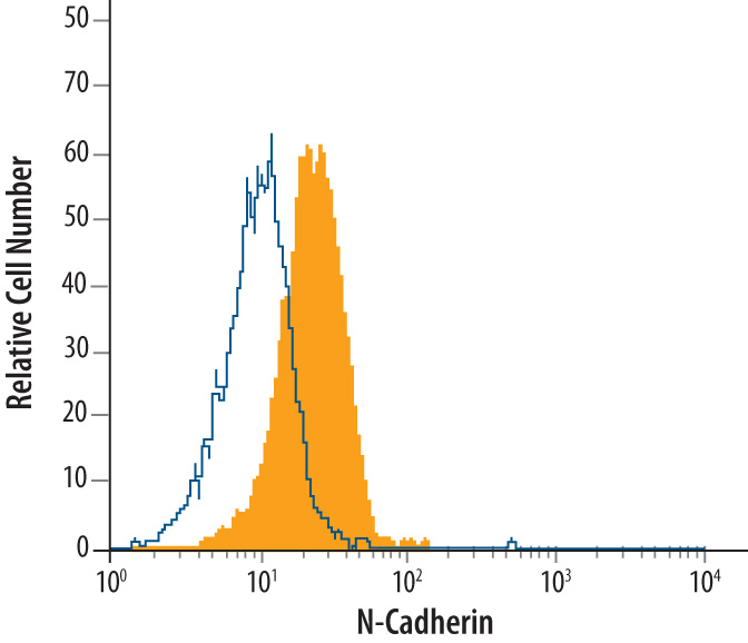
Detection of N‑Cadherin in HeLa Human Cell Line by Flow Cytometry. HeLa human cervical epithelial carcinoma cell line was stained with Sheep Anti-Human/Mouse/Rat N-Cadherin Antigen Affinity-purified Polyclonal Antibody (Catalog # AF6426, filled histogram) or Sheep IgG control antibody (Catalog # 5-001-A, open histogram), followed by NorthernLights™ 557-conjugated Anti-Sheep IgG Secondary Antibody (Catalog # NL010).
 View Larger
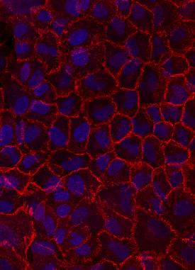
N‑Cadherin in A549 Human Cell Line. N-Cadherin was detected in immersion fixed A549 human lung carcinoma cell line using Sheep Anti-Human/Mouse/Rat N-Cadherin Affinity-purified Polyclonal Antibody (Catalog # AF6426) at 10 µg/mL for 3 hours at room temperature. Cells were stained using the NorthernLights™ 557-conjugated Anti-Sheep IgG Secondary Antibody (red; Catalog # NL010) and counterstained with DAPI (blue). Specific staining was localized to cell surfaces. View our protocol for Fluorescent ICC Staining of Cells on Coverslips.
 View Larger
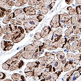
N‑Cadherin in Human Heart N-Cadherin was detected in immersion fixed paraffin-embedded sections of human heart using Sheep Anti-Human/Mouse/Rat N-Cadherin Antigen Affinity-purified Polyclonal Antibody (Catalog # AF6426) at 1 µg/mL overnight at 4 °C. Tissue was stained using the Sheep IgG VisUCyte HRP Polymer Antibody (Catalog # VC006). Before incubation with the primary antibody, tissue was subjected to heat-induced epitope retrieval using Antigen Retrieval Reagent-Basic (Catalog # CTS013). Tissue was stained using DAB (brown) and counterstained with hematoxylin (blue). Specific staining was localized to intercalated disks between the myocardial cells. Staining was performed using our protocol for IHC Staining with VisUCyte HRP Polymer Detection Reagents.
 View Larger
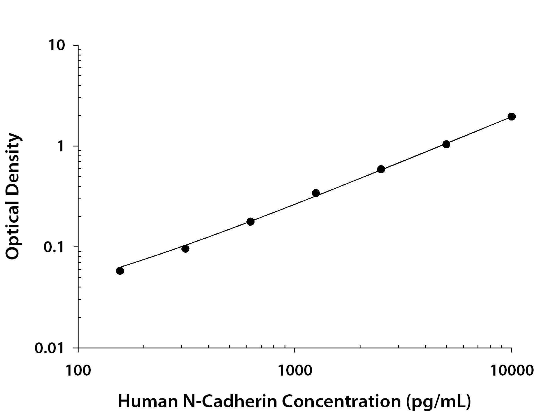
Human N‑Cadherin ELISA Standard Curve. Recombinant Human N-Cadherin protein was serially diluted 2-fold and captured by Mouse Anti-Human N-Cadherin Monoclonal Antibody (Catalog # MAB13883) coated on a Clear Polystyrene Microplate (Catalog # DY990). Sheep Anti-Human/Mouse/Rat N-Cadherin Antigen Affinity-purified Polyclonal Antibody (Catalog # AF6426) was biotinylated and incubated with the protein captured on the plate. Detection of the standard curve was achieved by incubating Streptavidin-HRP (Catalog # DY998) followed by Substrate Solution (Catalog # DY999) and stopping the enzymatic reaction with Stop Solution (Catalog # DY994).
Reconstitution Calculator
Preparation and Storage
- 12 months from date of receipt, -20 to -70 °C as supplied.
- 1 month, 2 to 8 °C under sterile conditions after reconstitution.
- 6 months, -20 to -70 °C under sterile conditions after reconstitution.
Background: N-Cadherin
N-Cadherin (Neural Cadherin; also CD325 and Cadherin-2) is a 130-135 kDa member of the "classical" (or type I) cadherin subfamily, cadherin superfamily of proteins. It is expressed on multiple cell types, including neurons, fibroblasts, Schwann cells, endothelial cells and hepatic stellate cells. N-Cadherin mediates homotypic binding, either in cis (same cell) or trans (adjacent cell). proN-Cadherin is expressed as an 881 amino acid (aa) type I transmembrane glycoprotein. It may be initially inserted into the ER, where the 15-20 kDa prodomain (aa 26-159) is cleaved by proprotein convertase, and the mature molecule (aa 160-906) is transported to the surface. Mature N-Cadherin contains a 565 aa extracellular region (aa 160-724) that possesses five cadherin domains (aa 160-714), and a 161 aa cytoplasmic tail that undergoes phosphorylation at Tyr785. There is one splice variant that contains a 10 aa substitution for aa 839-906. Over aa 160-724, human N Cadherin shares 98% aa identity with mouse N-Cadherin.
Product Datasheets
Citations for Human/Mouse/Rat N-Cadherin Antibody
R&D Systems personnel manually curate a database that contains references using R&D Systems products. The data collected includes not only links to publications in PubMed, but also provides information about sample types, species, and experimental conditions.
12
Citations: Showing 1 - 10
Filter your results:
Filter by:
-
An integrated organoid omics map extends modeling potential of kidney disease
Authors: Lassé, M;El Saghir, J;Berthier, CC;Eddy, S;Fischer, M;Laufer, SD;Kylies, D;Hutzfeldt, A;Bonin, LL;Dumoulin, B;Menon, R;Vega-Warner, V;Eichinger, F;Alakwaa, F;Fermin, D;Billing, AM;Minakawa, A;McCown, PJ;Rose, MP;Godfrey, B;Meister, E;Wiech, T;Noriega, M;Chrysopoulou, M;Brandts, P;Ju, W;Reinhard, L;Hoxha, E;Grahammer, F;Lindenmeyer, MT;Huber, TB;Schlüter, H;Thiel, S;Mariani, LH;Puelles, VG;Braun, F;Kretzler, M;Demir, F;Harder, JL;Rinschen, MM;
Nature communications
Species: Human
Sample Types: Whole Cells
Applications: Immunocytochemistry/ Immunofluorescence -
Integrating Oxygen and 3D Cell Culture System: A Simple Tool to Elucidate the Cell Fate Decision of hiPSCs
Authors: RR Khadim, RK Vadivelu, T Utami, FG Torizal, M Nishikawa, Y Sakai
International Journal of Molecular Sciences, 2022-06-30;23(13):.
Species: Human
Sample Types: Whole Cells
Applications: ICC -
Specific Binding of Novel SPION-Based System Bearing Anti-N-Cadherin Antibodies to Prostate Tumor Cells
Authors: K Karnas, T Str?czek, C Kapusta, M Lekka, J Duli?ska-L, A Karewicz
International Journal of Nanomedicine, 2021-09-24;16(0):6537-6552.
Species: Human
Sample Types: Whole Cells
Applications: Flow Cytometry -
Reciprocal interplay between asporin and decorin: Implications in gastric cancer prognosis
Authors: D Basak, Z Jamal, A Ghosh, PK Mondal, P Dey Talukd, S Ghosh, B Ghosh Roy, R Ghosh, A Halder, A Chowdhury, GK Dhali, BK Chattopadh, ML Saha, A Basu, S Roy, C Mukherjee, NK Biswas, U Chatterji, S Datta
PLoS ONE, 2021-08-11;16(8):e0255915.
Species: Human
Sample Types: Tissue Homogenates
Applications: Western Blot -
Inhibition of Sema4D/PlexinB1 signaling alleviates vascular dysfunction in diabetic retinopathy
Authors: JH Wu, YN Li, AQ Chen, CD Hong, CL Zhang, HL Wang, YF Zhou, PC Li, Y Wang, L Mao, YP Xia, QW He, HJ Jin, ZY Yue, B Hu
EMBO Mol Med, 2020-01-13;12(2):e10154.
Species: Mouse
Sample Types: Protein, Whole Cells
Applications: Functional Assay, Western Blot -
ADAM10 is Expressed by Ameloblasts, Cleaves the RELT TNF Receptor Extracellular Domain and Facilitates Enamel Development
Authors: A Ikeda, S Shahid, BR Blumberg, M Suzuki, JD Bartlett
Sci Rep, 2019-10-01;9(1):14086.
Species: Mouse
Sample Types: Protein
Applications: Western Blot -
Phenotypic heterogeneity and evolution of melanoma cells associated with targeted therapy resistance
Authors: Y Su, M Bintz, Y Yang, L Robert, AHC Ng, V Liu, A Ribas, JR Heath, W Wei
PLoS Comput. Biol., 2019-06-05;15(6):e1007034.
Species: Human
Sample Types: Whole Cells
Applications: ICC -
Organoid single cell profiling identifies a transcriptional signature of glomerular disease
Authors: JL Harder, R Menon, EA Otto, J Zhou, S Eddy, NL Wys, C O'Connor, J Luo, V Nair, C Cebrian, JR Spence, M Bitzer, OG Troyanskay, JB Hodgin, RC Wiggins, BS Freedman, M Kretzler
JCI Insight, 2019-01-10;4(1):.
Species: Human
Sample Types: Whole Tissue
Applications: IHC-P -
MicroRNA-149-5p regulates blood-brain barrier permeability after transient middle cerebral artery occlusion in rats by targeting S1PR2 of pericytes
Authors: Y Wan, HJ Jin, YY Zhu, Z Fang, L Mao, Q He, YP Xia, M Li, Y Li, X Chen, B Hu
FASEB J., 2018-01-18;0(0):fj201701121R.
Species: Rat
Sample Types: Cell Lysates, Whole Cells
Applications: ICC, Western Blot -
BDNF/TrkB axis activation promotes epithelial-mesenchymal transition in idiopathic pulmonary fibrosis
Authors: E Cherubini, S Mariotta, D Scozzi, R Mancini, G Osman, M D'Ascanio, P Bruno, G Cardillo, A Ricci
J Transl Med, 2017-09-22;15(1):196.
Species: Human
Sample Types: Whole Cells
Applications: ICC -
MMP20 modulates cadherin expression in ameloblasts as enamel develops.
Authors: Guan X, Bartlett J
2013-09-25;92(0):.
Species: Mouse
Sample Types: Cell Lysates
Applications: Western Blot -
In vivo biomarker expression patterns are preserved in 3D cultures of Prostate Cancer.
Authors: Windus L, Kiss D, Glover T, Avery V
Exp Cell Res, 2012-07-27;318(19):2507-19.
Species: Human
Sample Types: Cell Lysates, Whole Cells
Applications: ICC, Western Blot
FAQs
No product specific FAQs exist for this product, however you may
View all Antibody FAQsReviews for Human/Mouse/Rat N-Cadherin Antibody
There are currently no reviews for this product. Be the first to review Human/Mouse/Rat N-Cadherin Antibody and earn rewards!
Have you used Human/Mouse/Rat N-Cadherin Antibody?
Submit a review and receive an Amazon gift card.
$25/€18/£15/$25CAN/¥75 Yuan/¥2500 Yen for a review with an image
$10/€7/£6/$10 CAD/¥70 Yuan/¥1110 Yen for a review without an image


