Human/Mouse/Rat p53 Antibody Summary
Asp7-Asp393
Accession # P04637
Applications
Please Note: Optimal dilutions should be determined by each laboratory for each application. General Protocols are available in the Technical Information section on our website.
Scientific Data
 View Larger
Detection of Human p53 by Western Blot. Western blot shows lysates of CEM human T-lymphoblastoid cell line and MCF-7 human breast cancer cell line were mock-treated (-) or exposed (+) to 10 Gy ionizing radiation (IR) and harvested after 1 hour. PVDF membrane was probed with 0.5 µg/mL of Goat Anti-Human/Mouse/Rat p53 Antigen Affinity-purified Polyclonal Antibody (Catalog # AF1355), followed by HRP-conjugated Anti-Goat IgG Secondary Antibody (Catalog # HAF109). A specific band was detected for p53 at approximately 53 kDa (as indicated). This experiment was conducted under reducing conditions and using Immunoblot Buffer Group 1.
 View Larger
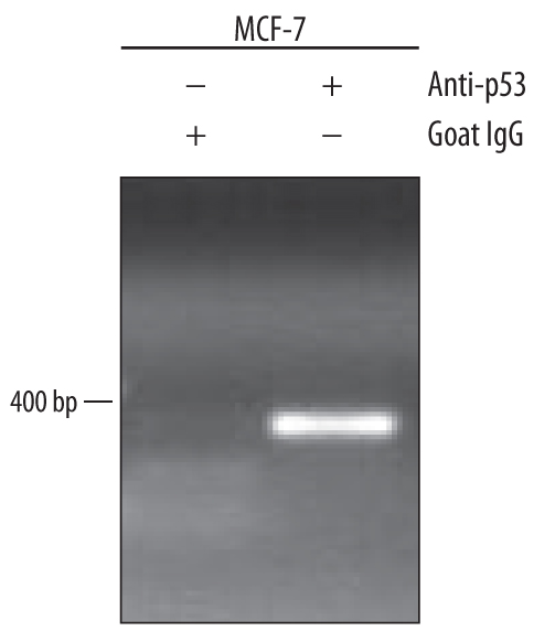
Detection of p53-regulated Genes by Chromatin Immunoprecipitation. MCF-7 human breast cancer cell line treated with 300 nM camptothecin overnight were fixed using formaldehyde, resuspended in lysis buffer, and sonicated to shear chromatin. p53/DNA complexes were immunoprecipitated using 5 µg Goat Anti-Human/Mouse/Rat p53 Antigen Affinity-purified Polyclonal Antibody (Catalog # AF1355) or control antibody (Catalog # AB-108-C) for 15 minutes in an ultrasonic bath, followed by Biotinylated Anti-Goat IgG Secondary Antibody (Catalog # BAF109). Immunocomplexes were captured using 50 µL of MagCellect Streptavidin Ferrofluid (Catalog # MAG999) and DNA was purified using chelating resin solution. The p21 promoter was detected by standard PCR.
 View Larger
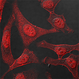
p53 in HeLa Human Cell Line. p53 was detected in immersion fixed HeLa human cervical epithelial carcinoma cell line using Goat Anti-Human/Mouse/Rat p53 Antigen Affinity-purified Polyclonal Antibody (Catalog # AF1355) at 1.7 µg/mL for 3 hours at room temperature. Cells were stained using the NorthernLights™ 557-conjugated Anti-Goat IgG Secondary Antibody (red; Catalog # NL001) and counterstained with DAPI (blue). Specific staining was localized to nuclei. View our protocol for Fluorescent ICC Staining of Cells on Coverslips.
 View Larger
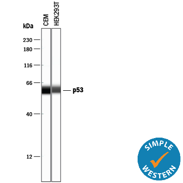
Detection of Human p53 by Simple WesternTM. Simple Western lane view shows lysates of CEM human T-lymphoblastoid cell line and HEK293T human embryonic kidney cell line, loaded at 0.2 mg/mL. A specific band was detected for p53 at approximately 59 kDa (as indicated) using 2.5 µg/mL of Goat Anti-Human/Mouse/Rat p53 Antigen Affinity-purified Polyclonal Antibody (Catalog # AF1355) followed by 1:50 dilution of HRP-conjugated Anti-Goat IgG Secondary Antibody (Catalog # HAF109). This experiment was conducted under reducing conditions and using the 12-230 kDa separation system.
 View Larger
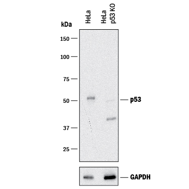
Western Blot Shows Human p53 Specificity by Using Knockout Cell Line. Western blot shows lysates of HeLa human cervical epithelial carcinoma parental cell line and p53 knockout HeLa cell line (KO). PVDF membrane was probed with 0.25 µg/mL of Goat Anti-Human/Mouse/Rat p53 Antigen Affinity-purified Polyclonal Antibody (Catalog # AF1355) followed by HRP-conjugated Anti-Goat IgG Secondary Antibody (Catalog # HAF017). A specific band was detected for p53 at approximately 53 kDa (as indicated) in the parental HeLa cell line, but is not detectable in knockout HeLa cell line. GAPDH (Catalog # AF5718) is shown as a loading control.This experiment was conducted under reducing conditions and using Immunoblot Buffer Group 1.
Reconstitution Calculator
Preparation and Storage
- 12 months from date of receipt, -20 to -70 °C as supplied.
- 1 month, 2 to 8 °C under sterile conditions after reconstitution.
- 6 months, -20 to -70 °C under sterile conditions after reconstitution.
Background: p53
The p53 tumor suppressor protein is a multi-functional transcription factor that regulates cellular decisions regarding proliferation, cell cycle checkpoints, and apoptosis. The importance of p53 is underscored by its mutation in over 50% of human cancers. Mice that lack one or both copies of p53 also showed an increased incidence of tumors, which makes the p53 deficient mouse a model system for studying cancer generation and progression.
Product Datasheets
Citations for Human/Mouse/Rat p53 Antibody
R&D Systems personnel manually curate a database that contains references using R&D Systems products. The data collected includes not only links to publications in PubMed, but also provides information about sample types, species, and experimental conditions.
15
Citations: Showing 1 - 10
Filter your results:
Filter by:
-
Calorie restriction alters the mechanisms of radiation-induced mouse thymic lymphomagenesis
Authors: T Nakayama, M Sunaoshi, Y Shang, M Takahashi, T Saito, BJ Blyth, Y Amasaki, K Daino, Y Shimada, A Tachibana, S Kakinuma
PLoS ONE, 2023-01-20;18(1):e0280560.
Species: Mouse
Sample Types: Protein
Applications: Western Blot -
Non-targeting control for MISSION shRNA library silences SNRPD3 leading to cell death or permanent growth arrest
Authors: Czarnek M, Sarad K, Kara? A et al.
Molecular Therapy - Nucleic Acids
-
Accumulation of cholesterol, triglycerides and ceramides in hepatocellular carcinomas of diethylnitrosamine injected mice
Authors: EM Haberl, R Pohl, L Rein-Fisch, M Höring, S Krautbauer, G Liebisch, C Buechler
Lipids in Health and Disease, 2021-10-10;20(1):135.
Species: Mouse
Sample Types: Tissue Homogenates
Applications: Western Blot -
MYC Promotes Bone Marrow Stem Cell Dysfunction in Fanconi Anemia
Authors: A Rodríguez, K Zhang, A Färkkilä, J Filiatraul, C Yang, M Velázquez, E Furutani, DC Goldman, B García de, G Garza-Mayé, K McQueen, LA Sambel, B Molina, L Torres, M González, E Vadillo, R Pelayo, WH Fleming, M Grompe, A Shimamura, S Hautaniemi, J Greenberge, S Frías, K Parmar, AD D'Andrea
Cell Stem Cell, 2020-09-29;0(0):.
Species: Human
Sample Types: Whole Cells
Applications: Flow Cytometry -
Germline mutation of MDM4, a major p53 regulator, in a familial syndrome of defective telomere maintenance
Authors: E Toufektcha, V Lejour, R Durand, N Giri, I Draskovic, B Bardot, P Laplante, S Jaber, BP Alter, JA Londono-Va, SA Savage, F Toledo
Sci Adv, 2020-04-10;6(15):eaay3511.
Species: Mouse
Sample Types: Cell Culture Lysates
Applications: Western Blot -
Progranulin protects the mouse retina under hypoxic conditions via inhibition of the Toll?like receptor?4?NADPH oxidase 4 signaling pathway
Authors: ZP You, MJ Yu, YL Zhang, K Shi
Mol Med Rep, 2018-11-08;0(0):.
Species: Mouse
Sample Types: Tissue Homogenates
Applications: Western Blot -
B-cell lymphoma 2 is associated with advanced tumor grade and clinical stage, and reduced overall survival in young Chinese patients with colorectal carcinoma
Authors: J Wang, G He, Q Yang, L Bai, B Jian, Q Li, Z Li
Oncol Lett, 2018-04-13;15(6):9009-9016.
Species: Human
Sample Types: Tissue Homogenates, Whole Tissue
Applications: IHC-P, Western Blot -
Chromosomal instability induced by increased BIRC5/Survivin levels affects tumorigenicity of glioma cells
Authors: M Conde, S Michen, R Wiedemuth, B Klink, E Schröck, G Schackert, A Temme
BMC Cancer, 2017-12-28;17(1):889.
Species: Human
Sample Types: Cell Lysates
Applications: Western Blot -
Dysfunction of the MDM2/p53 axis is linked to premature aging
Authors: D Lessel, D Wu, C Trujillo, T Ramezani, I Lessel, MK Alwasiyah, B Saha, FM Hisama, K Rading, I Goebel, P Schütz, G Speit, J Högel, H Thiele, G Nürnberg, P Nürnberg, M Hammerschm, Y Zhu, DR Tong, C Katz, GM Martin, J Oshima, C Prives, C Kubisch
J. Clin. Invest., 2017-08-28;0(0):.
Species: Human
Sample Types: Cell Lysates
Applications: Western Blot -
Combining anti-miR-155 with chemotherapy for the treatment of lung cancers
Authors: Katrien Van Roosbr
Clin. Cancer Res, 2016-11-30;0(0):.
Species: Human
Sample Types: Cell Lysates
Applications: Western Blot -
Growth hormone is permissive for neoplastic colon growth
Proc Natl Acad Sci USA, 2016-05-25;0(0):.
Species: Human
Sample Types: Cell Lysates
Applications: Western Blot -
Survivin safeguards chromosome numbers and protects from aneuploidy independently from p53.
Authors: Wiedemuth R, Klink B, Topfer K, Schrock E, Schackert G, Tatsuka M, Temme A
Mol Cancer, 2014-05-09;13(0):107.
Species: Human
Sample Types: Cell Lysates
Applications: Western Blot -
Expression of S100A6 in cardiac myocytes limits apoptosis induced by tumor necrosis factor-alpha.
Authors: Tsoporis JN, Izhar S, Parker TG
J. Biol. Chem., 2008-08-27;283(44):30174-83.
Species: Rat
Sample Types: Cell Lysates
Applications: Western Blot -
Protection of human keratinocytes from UVB-induced inflammation using root extract of Lithospermum erythrorhizon.
Authors: Ishida T, Sakaguchi I
Biol. Pharm. Bull., 2007-05-01;30(5):928-34.
Species: Human
Sample Types: Cell Lysates
Applications: Western Blot -
TIMP-1 gene deficiency increases tumour cell sensitivity to chemotherapy-induced apoptosis.
Authors: Davidsen ML, Wurtz SØ, Romer MU, Sorensen NM, Johansen SK, Christensen IJ, Larsen JK, Offenberg H, Brunner N, Lademann U
Br. J. Cancer, 2006-10-23;95(8):1114-20.
Species: Mouse
Sample Types: Cell Lysates
Applications: Western Blot
FAQs
No product specific FAQs exist for this product, however you may
View all Antibody FAQsReviews for Human/Mouse/Rat p53 Antibody
Average Rating: 5 (Based on 3 Reviews)
Have you used Human/Mouse/Rat p53 Antibody?
Submit a review and receive an Amazon gift card.
$25/€18/£15/$25CAN/¥75 Yuan/¥2500 Yen for a review with an image
$10/€7/£6/$10 CAD/¥70 Yuan/¥1110 Yen for a review without an image
Filter by:


