Human/Mouse/Rat Phospho-JNK (T183/Y185) Antibody Summary
Applications
Please Note: Optimal dilutions should be determined by each laboratory for each application. General Protocols are available in the Technical Information section on our website.
Scientific Data
 View Larger
Detection of Human and Mouse Phospho-JNK (T183/Y185) by Western Blot. Western blot shows lysates of HEK293 human embryonic kidney cell line and NIH-3T3 mouse embryonic fibroblast cell line untreated (-) or treated (+) with 100 J/m2UV-C for 30 minutes. PVDF membrane was probed with 0.5 µg/mL of Rabbit Anti-Human/Mouse/Rat Phospho-JNK (T183/Y185) Antigen Affinity-purified Polyclonal Antibody (Catalog # AF1205), followed by HRP-conjugated Anti-Rabbit IgG Secondary Antibody (Catalog # HAF008). Specific bands were detected for Phospho-JNK (T183/Y185) at approximately 46 and 54 kDa (as indicated). This experiment was conducted under reducing conditions and using Immunoblot Buffer Group 6.
 View Larger
JNK in Human Breast Cancer Tissue. JNK phosphorylated at T183/Y185 was detected in immersion fixed paraffin-embedded sections of human breast cancer tissue using Rabbit Anti-Human/Mouse/Rat Phospho-JNK (T183/Y185) Antigen Affinity-purified Polyclonal Antibody (Catalog # AF1205) at 15 µg/mL overnight at 4 °C. Tissue was stained using the Anti-Rabbit HRP-DAB Cell & Tissue Staining Kit (brown; Catalog # CTS005) and counterstained with hematoxylin (blue). View our protocol for Chromogenic IHC Staining of immersion fixed paraffin-embedded Tissue Sections.
 View Larger
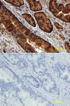
JNK in Human Prostate. JNK phosphorylated at T183/Y185 was detected in immersion fixed paraffin-embedded sections of human prostate array using Rabbit Anti-Human/Mouse/Rat Phospho-JNK (T183/Y185) Antigen Affinity-purified Polyclonal Antibody (Catalog # AF1205) at 15 µg/mL overnight at 4 °C. Tissue was stained using the Anti-Rabbit HRP-DAB Cell & Tissue Staining Kit (brown; Catalog # CTS005) and counterstained with hematoxylin (blue). Lower panel shows a lack of labeling if primary antibodies are omitted and tissue is stained only with secondary antibody followed by incubation with detection reagents. View our protocol for Chromogenic IHC Staining of Paraffin-embedded Tissue Sections.
 View Larger
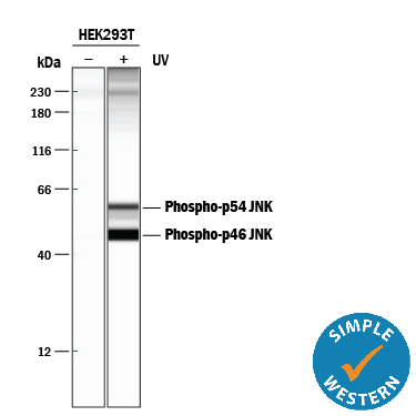
Detection of Human Phospho-JNK (T183/Y185) by Simple WesternTM. Simple Western lane view shows lysates of HEK293T human embryonic kidney cell line untreated (-) or treated (+) with 100 J/m2ultraviolet light (UV) for 30 minutes, loaded at 0.2 mg/mL. Specific bands were detected for Phospho-JNK (T183/Y185) at approximately 56 and 46 kDa (as indicated) using 5 µg/mL of Rabbit Anti-Human/Mouse/Rat Phospho-JNK (T183/Y185) Antigen Affinity-purified Polyclonal Antibody (Catalog # AF1205). This experiment was conducted under reducing conditions and using the 12-230 kDa separation system.*Non-specific interaction with the 230 kDa Simple Western standard may be seen with this antibody.
Reconstitution Calculator
Preparation and Storage
- 12 months from date of receipt, -20 to -70 °C as supplied.
- 1 month, 2 to 8 °C under sterile conditions after reconstitution.
- 6 months, -20 to -70 °C under sterile conditions after reconstitution.
Background: JNK
The c-Jun N-terminal Kinases (JNKs) are part of the MAPK (mitogen-activated protein kinase) system that transmits signals from the extracellular milieu to both the cytoplasm and nucleus of the cell. Following perturbation at the cell membrane, MEKKs/MAP3Ks are initially activated, followed by their activation of MKKs/MAP2Ks, and MKKs activation of MAPKs/MAP(1)Ks. There are three classes of MAPKs: ERKs, p38 Kinases and JNKs. JNKs are 45-55 kDa protein products of three genes which, through alternative splicing, generate up to 10 possible isoforms. The phosphorylation targets for MAPKs vary, but include p53, c-MYC, ATF2 and c-Jun, the latter molecule representing the namesake for the enzyme group. The three human JNKs share approximately 80% aa sequence identity. JNKs from human, mouse and rat all contain a conserved Met-Met-Thr(183)-Pro-Tyr(185)-Val-Val motif that undergoes dual phosphorylation by MMK4 and MMK7 to activate the different JNKs. Activated by environmental stresses and inflammatory cytokines, JNKs translocate to the nucleus where they regulate the activity of several transcription factors; including the c-Jun component of AP-1 and ATF-2.
Product Datasheets
Citations for Human/Mouse/Rat Phospho-JNK (T183/Y185) Antibody
R&D Systems personnel manually curate a database that contains references using R&D Systems products. The data collected includes not only links to publications in PubMed, but also provides information about sample types, species, and experimental conditions.
15
Citations: Showing 1 - 10
Filter your results:
Filter by:
-
Bag-1 Protects Nucleus Pulposus Cells from Oxidative Stress by Interacting with HSP70
Authors: K Suyama, D Sakai, S Hayashi, N Qu, H Terayama, D Kiyoshima, K Nagahori, M Watanabe
Biomedicines, 2023-03-12;11(3):.
Species: Human
Sample Types: Cell Lysates
Applications: Western Blot -
Changes of signaling molecules in the axotomized rat facial nucleus
Authors: T Ishijima, K Nakajima
Journal of chemical neuroanatomy, 2022-10-29;126(0):102179.
Species: Rat
Sample Types: Whole Tissue
Applications: IHC -
Novel molecular mechanisms underlying the ameliorative effect of N-acetyl-L-cysteine against ?-radiation-induced premature ovarian failure in rats
Authors: EM Mantawy, RS Said, DH Kassem, AK Abdel-Aziz, AM Badr
Ecotoxicol. Environ. Saf., 2020-08-29;206(0):111190.
Species: Rat
Sample Types: Whole Tissue
Applications: IHC -
Bim expression modulates the pro-inflammatory phenotype of retinal astroglial cells
Authors: J Falero-Per, N Sheibani, CM Sorenson
PLoS ONE, 2020-05-04;15(5):e0232779.
Species: Mouse
Sample Types: Cell Lysates
Applications: Western Blot -
Dysregulation of the splicing machinery is directly associated to aggressiveness of prostate cancer
Authors: JM Jiménez-Va, V Herrero-Ag, AJ Montero-Hi, E Gómez-Góme, AC Fuentes-Fa, AJ León-Gonzá, P Sáez-Martí, E Alors-Pére, S Pedraza-Ar, T González-S, O Reyes, A Martínez-L, R Sánchez-Sá, S Ventura, EM Yubero-Ser, MJ Requena-Ta, JP Castaño, MD Gahete, RM Luque
EBioMedicine, 2020-01-03;51(0):102547.
Species: Human
Sample Types:
Applications: Western Blot -
Effects of interleukin-17A in nucleus pulposus cells and its small-molecule inhibitors for intervertebral disc disease
Authors: K Suyama, D Sakai, N Hirayama, Y Nakamura, E Matsushita, H Terayama, N Qu, O Tanaka, K Sakabe, M Watanabe
J. Cell. Mol. Med., 2018-09-11;0(0):.
Species: Rat
Sample Types: Cell Lysates
Applications: Western Blot -
WWOX sensitises ovarian cancer cells to paclitaxel via modulation of the ER stress response
Authors: S Janczar, J Nautiyal, Y Xiao, E Curry, M Sun, E Zanini, AJ Paige, H Gabra
Cell Death Dis, 2017-07-27;8(7):e2955.
Species: Human
Sample Types: Cell Lysates
Applications: Western Blot -
Mechanistic clues to the protective effect of chrysin against doxorubicin-induced cardiomyopathy: Plausible roles of p53, MAPK and AKT pathways
Authors: EM Mantawy, A Esmat, WM El-Bakly, RA Salah ElDi, E El-Demerda
Sci Rep, 2017-07-06;7(1):4795.
Species: Rat
Sample Types: Tissue Homogenates
Applications: Western Blot -
White-to-brown metabolic conversion of human adipocytes by JAK inhibition.
Authors: Moisan A, Lee Y, Zhang J, Hudak C, Meyer C, Prummer M, Zoffmann S, Truong H, Ebeling M, Kiialainen A, Gerard R, Xia F, Schinzel R, Amrein K, Cowan C
Nat Cell Biol, 2014-12-08;17(1):57-67.
Species: Human
Sample Types: Cell Lysates
Applications: Western Blot -
High glucose alters retinal astrocytes phenotype through increased production of inflammatory cytokines and oxidative stress.
Authors: Shin E, Huang Q, Gurel Z, Sorenson C, Sheibani N
PLoS ONE, 2014-07-28;9(7):e103148.
Species: Mouse
Sample Types: Cell Lysates
Applications: Western Blot -
A p38MAPK/MK2 signaling pathway leading to redox stress, cell death and ischemia/reperfusion injury.
Authors: Ashraf M, Ebner M, Wallner C, Haller M, Khalid S, Schwelberger H, Koziel K, Enthammer M, Hermann M, Sickinger S, Soleiman A, Steger C, Vallant S, Sucher R, Brandacher G, Santer P, Dragun D, Troppmair J
Cell Commun Signal, 2014-01-14;12(0):6.
Species: Rat
Sample Types: Tissue Homogenates
Applications: Western Blot -
ERK5 protein promotes, whereas MEK1 protein differentially regulates, the Toll-like receptor 2 protein-dependent activation of human endothelial cells and monocytes.
Authors: Wilhelmsen K, Mesa K, Lucero J, Xu F, Hellman J
J Biol Chem, 2012-06-15;287(32):26478-94.
Species: Human
Sample Types: Cell Lysates
Applications: Western Blot -
Protective effect of the poly(ADP-ribose) polymerase inhibitor PJ34 on mitochondrial depolarization-mediated cell death in hepatocellular carcinoma cells involves attenuation of c-Jun N-terminal kinase-2 and protein kinase B/Akt activation.
Mol. Cancer, 2012-05-14;11(0):34.
Species: Human
Sample Types: Cell Lysates
Applications: Western Blot -
Oxidized alpha1-antitrypsin stimulates the release of monocyte chemotactic protein-1 from lung epithelial cells: potential role in emphysema.
Authors: Li Z, Alam S, Wang J, Sandstrom CS, Janciauskiene S, Mahadeva R
Am. J. Physiol. Lung Cell Mol. Physiol., 2009-06-12;297(2):L388-400.
Species: Human
Sample Types: Cell Lysates
Applications: Western Blot -
Common dysregulation of Wnt/Frizzled receptor elements in human hepatocellular carcinoma.
Authors: Bengochea A, de Souza MM, Lefrancois L, Le Roux E, Galy O, Chemin I, Kim M, Wands JR, Trepo C, Hainaut P, Scoazec JY, Vitvitski L, Merle P
Br. J. Cancer, 2008-06-24;99(1):143-50.
Species: Human
Sample Types: Cell Lysates
Applications: Western Blot
FAQs
No product specific FAQs exist for this product, however you may
View all Antibody FAQsReviews for Human/Mouse/Rat Phospho-JNK (T183/Y185) Antibody
Average Rating: 3.7 (Based on 3 Reviews)
Have you used Human/Mouse/Rat Phospho-JNK (T183/Y185) Antibody?
Submit a review and receive an Amazon gift card.
$25/€18/£15/$25CAN/¥75 Yuan/¥2500 Yen for a review with an image
$10/€7/£6/$10 CAD/¥70 Yuan/¥1110 Yen for a review without an image
Filter by:


