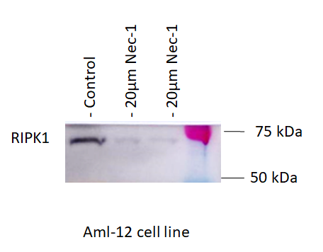
Human/Mouse/Rat RIPK1/RIP1 Antibody Summary
Met1-Asn671
Accession # Q13546
Applications
Please Note: Optimal dilutions should be determined by each laboratory for each application. General Protocols are available in the Technical Information section on our website.
Scientific Data
 View Larger
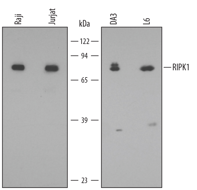
Detection of Human/Mouse/Rat RIPK1/RIP1 by Western Blot. Western blot shows lysates of Raji human Burkitt's lymphoma cell line, Jurkat human acute T cell leukemia cell line, DA3 mouse myeloma cell line, and L6 rat myoblast cell line. PVDF membrane was probed with 0.5 µg/mL of Mouse Anti-Human/Mouse/Rat RIPK1/RIP1 Monoclonal Antibody (Catalog # MAB3585) followed by HRP-conjugated Anti-Mouse IgG Secondary Antibody (Catalog # HAF007). A specific band was detected for RIPK1/RIP1 at approximately 75 kDa (as indicated). This experiment was conducted under reducing conditions and using Immunoblot Buffer Group 2.
 View Larger
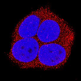
RIPK1/RIP1 in MCF‑7 Human Cell Line. RIPK1/RIP1 was detected in immersion fixed MCF-7 human breast cancer cell line using Mouse Anti-Human/Mouse/Rat RIPK1/RIP1 Monoclonal Antibody (Catalog # MAB3585) at 25 µg/mL for 3 hours at room temperature. Cells were stained using the NorthernLights™ 557-conjugated Anti-Mouse IgG Secondary Antibody (red; Catalog # NL007) and counterstained with DAPI (blue). Specific staining was localized to cytoplasm. View our protocol for Fluorescent ICC Staining of Cells on Coverslips.
 View Larger
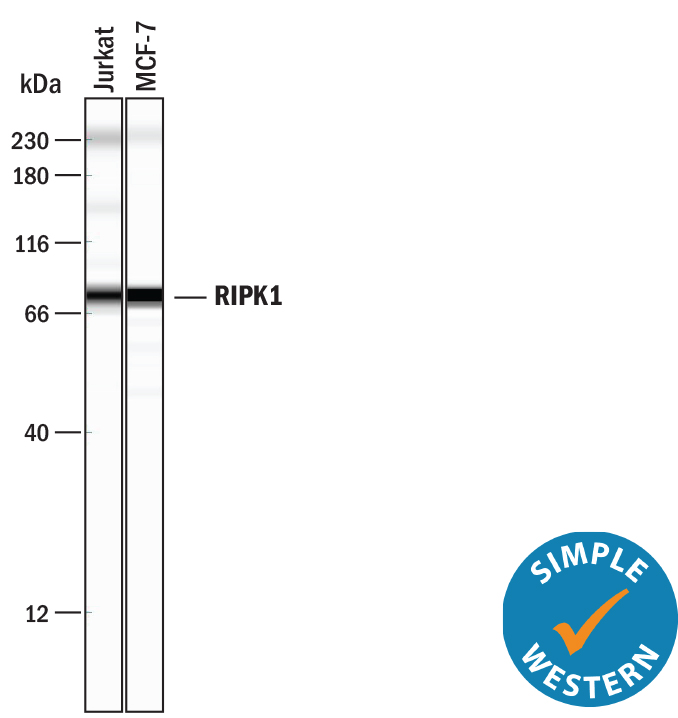
Detection of Human RIPK1/RIP1 by Simple WesternTM. Simple Western lane view shows lysates of Jurkat human acute T cell leukemia cell line and MCF‑7 human breast cancer cell line, loaded at 0.2 mg/mL. A specific band was detected for RIPK1/RIP1 at approximately 78 kDa (as indicated) using 10 µg/mL of Mouse Anti-Human/Mouse/Rat RIPK1/RIP1 Monoclonal Antibody (Catalog # MAB3585). This experiment was conducted under reducing conditions and using the 12-230 kDa separation system.
Non-specific interaction with the 230 kDa Simple Western standard may be seen with this antibody.
 View Larger
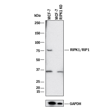
Western Blot Shows Human RIPK1/RIP1 Specificity by Using Knockout Cell Line. Western blot shows lysates of MCF-7 human breast cancer parental cell line and RIPK1/RIP1 knockout MCF-7 cell line (KO). PVDF membrane was probed with 0.5 µg/mL of Mouse Anti-Human/Mouse/Rat RIPK1/RIP1 Monoclonal Antibody (Catalog # MAB3585) followed by HRP-conjugated Anti-Mouse IgG Secondary Antibody (Catalog # HAF018). A specific band was detected for RIPK1/RIP1 at approximately 75 kDa (as indicated) in the parental MCF-7 cell line, but is not detectable in knockout MCF-7 cell line. GAPDH (Catalog # MAB5718) is shown as a loading control. This experiment was conducted under reducing conditions and using Immunoblot Buffer Group 1.
Reconstitution Calculator
Preparation and Storage
- 12 months from date of receipt, -20 to -70 °C as supplied.
- 1 month, 2 to 8 °C under sterile conditions after reconstitution.
- 6 months, -20 to -70 °C under sterile conditions after reconstitution.
Background: RIPK1/RIP1
Receptor-Interacting Protein 1 (RIP1, also known as RIPK1) is a 671 amino acid (aa) 75 kDa protein that contains an N-terminal protein kinase domain, a C-terminal death domain, and a unique internal region called the intermediate domain. RIP1 is a serine/threonine protein kinase and is constitutively expressed in many tissues. RIP1 interacts with the cytoplasmic death domain of FAS and TNF receptors and is an important element in the signal transduction machinery that mediates apoptosis. RIP1 has been shown to interact with a number of proteins including TRADD, TRAF1, TRAF2, and TRAF3, to form larger signaling complexes. These complexes, in turn, activate specific signaling cascades, such as NF kappa B. RIP1 also interacts through the C-terminal RIP homotypic interaction motif (RHIM) of TRIF in TLR3 dependent activation of NF kappa B.
Product Datasheets
Citations for Human/Mouse/Rat RIPK1/RIP1 Antibody
R&D Systems personnel manually curate a database that contains references using R&D Systems products. The data collected includes not only links to publications in PubMed, but also provides information about sample types, species, and experimental conditions.
13
Citations: Showing 1 - 10
Filter your results:
Filter by:
-
Anagliptin prevents lipopolysaccharide (LPS)- induced inflammation and activation of macrophages
Authors: F Yu, W Tian, J Dong
International immunopharmacology, 2022-01-16;104(0):108514.
Species: Mouse
Sample Types: Cell Lysates
Applications: Western Blot -
A Novel Method of Mouse RPE Explant Culture and Effective Introduction of Transgenes Using Adenoviral Transduction for In Vitro Studies in AMD
Authors: P Shang, NA Stepicheva, H Liu, O Chowdhury, J Franks, M Sun, S Hose, S Ghosh, M Yazdankhah, A Strizhakov, DB Stolz, JS Zigler, D Sinha
International Journal of Molecular Sciences, 2021-11-05;22(21):.
Species: Mouse
Sample Types: Cell Lysates
Applications: Western Blot -
ABT?737, a Bcl?2 family inhibitor, has a synergistic effect with apoptosis by inducing urothelial carcinoma cell necroptosis
Authors: R Cheng, X Liu, Z Wang, K Tang
Molecular Medicine Reports, 2021-03-31;23(6):.
Species: Human
Sample Types: Cell Lysates
Applications: Western Blot -
Inhibition of MLKL Attenuates Necroptotic Cell Death in a Murine Cell Model of Ischaemia Injury
Authors: R Baidya, J Gautheron, DHG Crawford, H Wang, KR Bridle
Journal of Clinical Medicine, 2021-01-08;10(2):.
Species: Mouse
Sample Types: Cell Lysates, Whole Cells
Applications: Flow Cytometry, Western Blot -
Interaction between the BAG1S isoform and HSP70 mediates the stability of anti-apoptotic proteins and the survival of osteosarcoma cells expressing oncogenic MYC
Authors: VJ Gennaro, H Wedegaertn, SB McMahon
BMC Cancer, 2019-03-22;19(1):258.
Species: Human
Sample Types: Cell Lysates
Applications: Western Blot -
Leishmania braziliensis Subverts Necroptosis by Modulating RIPK3 Expression
Authors: NF Luz, R Khouri, J Van Weyenb, DL Zanette, PP Fiuza, A Noronha, A Barral, VS Boaventura, DB Prates, FK Chan, BB Andrade, VM Borges
Front Microbiol, 2018-09-28;9(0):2283.
Species: Human
Sample Types: Cell Lysates
Applications: Western Blot -
RIP3 dependent NLRP3 inflammasome activation is implicated in acute lung injury in mice
Authors: J Chen, S Wang, R Fu, M Zhou, T Zhang, W Pan, N Yang, Y Huang
J Transl Med, 2018-08-20;16(1):233.
Species: Human
Sample Types: Cell Lysates
Applications: Western Blot -
Necrostatin-1 protects C2C12 myotubes from CoCl2-induced hypoxia
Authors: R Chen, J Xu, Y She, T Jiang, S Zhou, H Shi, C Li
Int. J. Mol. Med., 2018-02-06;41(5):2565-2572.
Species: Mouse
Sample Types: Cell Lysates
Applications: Western Blot -
MPP+ induces necrostatin-1- and ferrostatin-1-sensitive necrotic death of neuronal SH-SY5Y cells
Authors: K Ito, Y Eguchi, Y Imagawa, S Akai, H Mochizuki, Y Tsujimoto
Cell Death Discov, 2017-02-27;3(0):17013.
Species: Human
Sample Types: Cell Lysates
Applications: Western Blot -
Hypoxia potentiates the cytotoxic effect of piperlongumine in pheochromocytoma models
Oncotarget, 2016-06-28;7(26):40531-40545.
Species: Mouse
Sample Types: Cell Lysates
Applications: Western Blot -
ESCRT-0 dysfunction compromises autophagic degradation of protein aggregates and facilitates ER stress-mediated neurodegeneration via apoptotic and necroptotic pathways
Authors: R Oshima, T Hasegawa, K Tamai, N Sugeno, S Yoshida, J Kobayashi, A Kikuchi, T Baba, A Futatsugi, I Sato, K Satoh, A Takeda, M Aoki, N Tanaka
Sci Rep, 2016-04-26;6(0):24997.
Species: Rat
Sample Types: Cell Lysates
Applications: Western Blot -
Open reading frame 3 of genotype 1 hepatitis E virus inhibits nuclear factor-kappaappa B signaling induced by tumor necrosis factor-alpha in human A549 lung epithelial cells.
Authors: Xu J, Wu F, Tian D, Wang J, Zheng Z, Xia N
PLoS ONE, 2014-06-24;9(6):e100787.
Species: Human
Sample Types: Cell Lysates
Applications: Western Blot -
TWEAK Induces Apoptosis through a Death-signaling Complex Comprising Receptor-interacting Protein 1 (RIP1), Fas-associated Death Domain (FADD), and Caspase-8.
Authors: Ikner A, Ashkenazi A
J. Biol. Chem., 2011-04-27;286(24):21546-54.
Species: Human
Sample Types: Cell Lysates
Applications: Western Blot
FAQs
No product specific FAQs exist for this product, however you may
View all Antibody FAQsReviews for Human/Mouse/Rat RIPK1/RIP1 Antibody
Average Rating: 5 (Based on 1 Review)
Have you used Human/Mouse/Rat RIPK1/RIP1 Antibody?
Submit a review and receive an Amazon gift card.
$25/€18/£15/$25CAN/¥75 Yuan/¥2500 Yen for a review with an image
$10/€7/£6/$10 CAD/¥70 Yuan/¥1110 Yen for a review without an image
Filter by:
