Human/Mouse/Rat TDP-43/TARDBP Antibody Summary
Met1-Thr103
Accession # Q13148
Applications
Please Note: Optimal dilutions should be determined by each laboratory for each application. General Protocols are available in the Technical Information section on our website.
Scientific Data
 View Larger
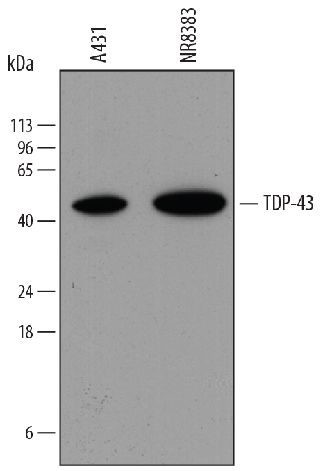
Detection of Human TDP-43/TARDBP by Western Blot. Western blot shows lysates of A431 human epithelial carcinoma cell line and NR8383 rat alveolar macrophage cell line. PVDF membrane was probed with 0.4 µg/mL of Mouse Anti-Human/Mouse/Rat TDP-43/TARDBP Monoclonal Antibody (Catalog # MAB7778) followed by HRP-conjugated Anti-Mouse IgG Secondary Antibody (HAF018). A specific band was detected for TDP-43/TARDBP at approximately 45 kDa (as indicated). This experiment was conducted under reducing conditions and using Immunoblot Buffer Group 1.
 View Larger
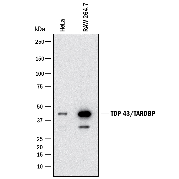
Detection of Human and Mouse TDP-43/TARDBP by Western Blot. Western blot shows lysates of HeLa human cervical epithelial carcinoma cell line and RAW 264.7 mouse monocyte/macrophage cell line. PVDF membrane was probed with 1 µg/mL of Mouse Anti-Human TDP-43/TARDBP Monoclonal Antibody (Catalog # MAB7778) followed by HRP-conjugated Anti-Mouse IgG Secondary Antibody (HAF018). A specific band was detected for TDP-43/TARDBP at approximately 43 kDa (as indicated). This experiment was conducted under reducing conditions and using Immunoblot Buffer Group 3.
 View Larger
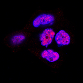
TDP-43/TARDBP in A431 Human Cell Line. TDP-43/TARDBP was detected in immersion fixed A431 human epithelial carcinoma cell line using Mouse Anti-Human/Mouse/Rat TDP-43/TARDBP Monoclonal Antibody (Catalog # MAB7778) at 1 µg/mL for 3 hours at room temperature. Cells were stained using the NorthernLights™ 557-conjugated Anti-Sheep IgG Secondary Antibody (red; NL010) and counterstained with DAPI (blue). Specific staining was localized to nuclei. View our protocol for Fluorescent ICC Staining of Cells on Coverslips.
 View Larger
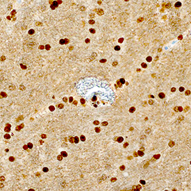
TDP-43/TARDBP in Human Brain. TDP-43/TARDBP was detected in immersion fixed paraffin-embedded sections of human brain (hippocampus) using Mouse Anti-Human/Mouse/Rat TDP-43/TARDBP Monoclonal Antibody (Catalog # MAB7778) at 1.7 µg/mL overnight at 4 °C. Tissue was stained using the Anti-Mouse HRP-DAB Cell & Tissue Staining Kit (brown; CTS002) and counterstained with hematoxylin (blue). Specific staining was localized to nuclei. View our protocol for Chromogenic IHC Staining of Paraffin-embedded Tissue Sections.
 View Larger
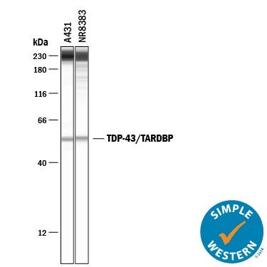
Detection of Human and Rat TDP-43/TARDBP by Simple WesternTM. Simple Western lane view shows lysates of A431 human epithelial carcinoma cell line and NR8383 rat alveolar macrophage cell line, loaded at 0.2 mg/mL. A specific band was detected for TDP-43/TARDBP at approximately 54 kDa (as indicated) using 5 µg/mL of Mouse Anti-Human TDP-43/TARDBP Monoclonal Antibody (Catalog # MAB7778). This experiment was conducted under reducing conditions and using the 12-230 kDa separation system. Non-specific interaction with the 230 kDa Simple Western standard may be seen with this antibody.
 View Larger
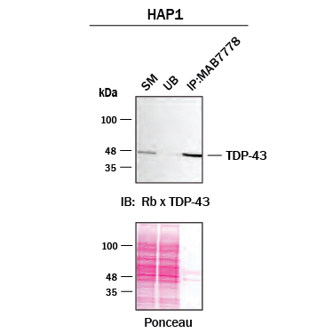
Detection of TDP-43/TARDBP by Immunoprecipitation. Immunoprecipitation was performed on cell lysate of HAP1 human near-haploid cell line using 2.0 μg of Mouse Anti-Human TDP-43 Monoclonal Antibody (Catalog # MAB7778) pre-coupled to protein G or protein A beads. Immunoprecipitated TDP-43/TARDBP was detected with a Rabbit Anti-TDP-43 antibody. The Ponceau stained transfers of each blot are shown. SM=10% starting material; UB=10% unbound fraction; IP=immunoprecipitated. Image, protocol, and testing courtesy of YCharOS Inc. (ycharos.com).
Reconstitution Calculator
Preparation and Storage
- 12 months from date of receipt, -20 to -70 °C as supplied.
- 1 month, 2 to 8 °C under sterile conditions after reconstitution.
- 6 months, -20 to -70 °C under sterile conditions after reconstitution.
Background: TDP-43/TARDBP
TDP-43 (TAR DNA binding protein of 43 kDa; gene name TARDBP) is a 414 amino acid (aa) nuclear RNA/DNA binding phosphoprotein that regulates transcription and splicing. TDP-43 is aberrantly expressed in most forms of amylotrophic lateral sclerosis and frontotemporal lobar degeneration. In these cases it is absent from the nucleus and accumulates, along with a truncated 20-28 kDa form, in ubiquitin-containing inclusion bodies. TDP-43 can also influence CFTR exon 9 skipping in cystic fibrosis. Within aa 1-103, human and mouse TDP-43 share 100% aa sequence identity, and 99% aa identity with rat TDP-43.
Product Datasheets
Citation for Human/Mouse/Rat TDP-43/TARDBP Antibody
R&D Systems personnel manually curate a database that contains references using R&D Systems products. The data collected includes not only links to publications in PubMed, but also provides information about sample types, species, and experimental conditions.
1 Citation: Showing 1 - 1
-
Epithelial-derived factors induce muscularis mucosa of human induced pluripotent stem cell-derived gastric organoids
Authors: K Uehara, M Koyanagi-A, T Koide, T Itoh, T Aoi
Stem Cell Reports, 2022-03-03;0(0):.
Species: Mouse
Sample Types: Tissue Lysates
Applications: Western Blot
FAQs
No product specific FAQs exist for this product, however you may
View all Antibody FAQsReviews for Human/Mouse/Rat TDP-43/TARDBP Antibody
Average Rating: 5 (Based on 1 Review)
Have you used Human/Mouse/Rat TDP-43/TARDBP Antibody?
Submit a review and receive an Amazon gift card.
$25/€18/£15/$25CAN/¥75 Yuan/¥2500 Yen for a review with an image
$10/€7/£6/$10 CAD/¥70 Yuan/¥1110 Yen for a review without an image
Filter by:


