Human/Mouse/Rat XIAP Antibody Summary
Met1-Ser497 (Ser162Cys)
Accession # P98170
Applications
Please Note: Optimal dilutions should be determined by each laboratory for each application. General Protocols are available in the Technical Information section on our website.
Scientific Data
 View Larger
XIAP in Human Lymphoma. XIAP was detected in immersion fixed paraffin-embedded sections of human lymphoma using 5 µg/mL Goat Anti-Human/Mouse/Rat XIAP Antigen Affinity-purified Polyclonal Antibody (Catalog # AF8221) overnight at 4 °C. Tissue was stained with the Anti-Goat HRP-DAB Cell & Tissue Staining Kit (brown; Catalog # CTS008) and counterstained with hematoxylin (blue). View our protocol for Chromogenic IHC Staining of Paraffin-embedded Tissue Sections.
 View Larger
XIAP in Human Breast Cancer Tissue. XIAP was detected in immersion fixed paraffin-embedded sections of human breast cancer tissue using 15 µg/mL Goat Anti-Human/Mouse/Rat XIAP Antigen Affinity-purified Polyclonal Antibody (Catalog # AF8221) overnight at 4 °C. Tissue was stained with the Anti-Goat HRP-DAB Cell & Tissue Staining Kit (brown; Catalog # CTS008) and counterstained with hematoxylin (blue). View our protocol for Chromogenic IHC Staining of Paraffin-embedded Tissue Sections.
 View Larger
Detection of Human/Mouse/Rat XIAP by Western Blot. Western blot shows lysates of Jurkat human acute T cell leukemia cell line, C2C12 mouse myoblast cell line, and PC-12 rat adrenal pheochromocytoma cell line. PVDF membrane was probed with 0.5 µg/mL of Goat Anti-Human/Mouse/Rat XIAP Antigen Affinity-purified Polyclonal Antibody (Catalog # AF8221) followed by HRP-conjugated Anti-Goat IgG Secondary Antibody (Catalog # HAF109). A specific band was detected for XIAP at approximately 56 kDa (as indicated). This experiment was conducted under reducing conditions and using Immunoblot Buffer Group 4.
 View Larger
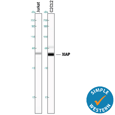
Detection of Human and Mouse XIAP by Simple WesternTM. Simple Western lane view shows lysates of Jurkat human acute T cell leukemia cell line and C2C12 mouse myoblast cell line, loaded at 0.2 mg/mL. A specific band was detected for XIAP at approximately 59 kDa (as indicated) using 5 µg/mL of Goat Anti-Human/Mouse/Rat XIAP Antigen Affinity-purified Polyclonal Antibody (Catalog # AF8221) followed by 1:50 dilution of HRP-conjugated Anti-Goat IgG Secondary Antibody (Catalog # HAF109). This experiment was conducted under reducing conditions and using the 12-230 kDa separation system.
 View Larger
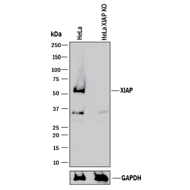
Western Blot Shows Human XIAP Specificity by Using Knockout Cell Line. Western blot shows lysates of HeLa human cervical epithelial carcinoma parental cell line and XIAP knockout HeLa cell line (KO). PVDF membrane was probed with 0.5 µg/mL of Goat Anti-Human/Mouse/Rat XIAP Antigen Affinity-purified Polyclonal Antibody (Catalog # AF8221) followed by HRP-conjugated Anti-Goat IgG Secondary Antibody (Catalog # HAF017). A specific band was detected for XIAP at approximately 52 kDa (as indicated) in the parental HeLa cell line, but is not detectable in knockout HeLa cell line. GAPDH (Catalog # AF5718) is shown as a loading control. This experiment was conducted under reducing conditions and using Immunoblot Buffer Group 1.
Reconstitution Calculator
Preparation and Storage
- 12 months from date of receipt, -20 to -70 °C as supplied.
- 1 month, 2 to 8 °C under sterile conditions after reconstitution.
- 6 months, -20 to -70 °C under sterile conditions after reconstitution.
Background: XIAP
XIAP (X-chromosome linked inhibitor of apoptosis) is a member of the apoptosis (IAP) family of proteins that inhibit caspases. The BIR2 domain of XIAP inhibits caspase-3 and caspase-7. The ability of XIAP to inhibit caspases is prevented by SMAC/Diablo through binding to XIAP-BIR2 and -BIR3 domains.
Product Datasheets
Citations for Human/Mouse/Rat XIAP Antibody
R&D Systems personnel manually curate a database that contains references using R&D Systems products. The data collected includes not only links to publications in PubMed, but also provides information about sample types, species, and experimental conditions.
16
Citations: Showing 1 - 10
Filter your results:
Filter by:
-
cIAP1/TRAF2 interplay promotes tumor growth through the activation of STAT3
Authors: B Dumétier, A Zadoroznyj, J Berthelet, S Causse, J Allègre, P Bourgeois, F Cattin, C Racoeur, C Paul, C Garrido, L Dubrez
Oncogene, 2022-11-18;0(0):.
Species: Human
Sample Types: Cell Lysates
Applications: Western Blot -
Nociceptor-derived Reg3gamma prevents endotoxic death by targeting kynurenine pathway in microglia
Authors: E Sugisawa, T Kondo, Y Kumagai, H Kato, Y Takayama, K Isohashi, E Shimosegaw, N Takemura, Y Hayashi, T Sasaki, MM Martino, M Tominaga, K Maruyama
Cell Reports, 2022-03-08;38(10):110462.
Species: Mouse
Sample Types: Cell Lysates
Applications: Western Blot -
Death agonist antibody against TRAILR2/DR5/TNFRSF10B enhances birinapant anti-tumor activity in HPV-positive head and neck squamous cell carcinomas
Authors: Y An, J Jeon, L Sun, A Derakhshan, J Chen, S Carlson, H Cheng, C Silvin, X Yang, C Van Waes, Z Chen
Scientific Reports, 2021-03-18;11(1):6392.
Species: Human
Sample Types: Cell Lysates
Applications: Western Blot -
ASTX660, a novel non-peptidomimetic antagonist of cIAP1/2 and XIAP, potently induces TNF-? dependent apoptosis in cancer cell lines and inhibits tumor growth
Authors: GA Ward, EJ Lewis, JS Ahn, CN Johnson, JF Lyons, V Martins, JM Munck, SJ Rich, T Smyth, NT Thompson, PA Williams, NE Wilsher, NG Wallis, G Chessari
Mol. Cancer Ther., 2018-04-25;0(0):.
Species: Human
Sample Types: Cell Lysates
Applications: Immunoprecipitation, Western Blot -
Chronic oxycodone induces axonal degeneration in rat brain
Authors: R Fan, LM Schrott, T Arnold, S Snelling, M Rao, D Graham, A Cornelius, NL Korneeva
BMC Neurosci, 2018-03-23;19(1):15.
Species: Rat
Sample Types: Tissue Homogenates
Applications: Western Blot -
Anticancer efficacy of the hypoxia-activated prodrug evofosfamide is enhanced in combination with proapoptotic receptor agonists against osteosarcoma
Authors: V Liapis, A Zysk, M DeNichilo, I Zinonos, S Hay, V Panagopoul, A Shoubridge, C Difelice, V Ponomarev, W Ingman, GJ Atkins, DM Findlay, ACW Zannettino, A Evdokiou
Cancer Med, 2017-08-10;0(0):.
Species: Human
Sample Types: Cell Lysates
Applications: Western Blot -
Delayed apoptosis allows islet ?-cells to implement an autophagic mechanism to promote cell survival
Authors: HL Hayes, BS Peterson, JM Haldeman, CB Newgard, HE Hohmeier, SB Stephens
PLoS ONE, 2017-02-17;12(2):e0172567.
Species: Rat
Sample Types: Cell Lysates
Applications: Western Blot -
Pro-Apoptotic Activity of New Honokiol/Triphenylmethane Analogues in B-Cell Lymphoid Malignancies
Molecules, 2016-07-30;21(8):.
Species: Human
Sample Types: Whole Cells
Applications: Flow Cytometry -
Delivery of a survivin promoter-driven antisense survivin-expressing plasmid DNA as a cancer therapeutic: a proof-of-concept study
Onco Targets Ther, 2016-05-03;9(0):2601-13.
Species: Human
Sample Types: Cell Lysates
Applications: Western Blot -
Doxorubicin overcomes resistance to drozitumab by antagonizing Inhibitor of Apoptosis Proteins (IAPs).
Authors: Zinonos I, Labrinidis A, Liapis V, Hay S, Panagopoulos V, Denichilo M, Ponomarev V, Ingman W, Atkins G, Findlay D, Zannettino A, Evdokiou A
Anticancer Res, 2014-12-01;34(12):7007-20.
Species: Human
Sample Types: Cell Lysates
Applications: Western Blot -
Cytotoxic activity of the amphibian ribonucleases onconase and r-amphinase on tumor cells from B cell lymphoproliferative disorders.
Authors: Smolewski P, Witkowska M, Zwolinska M, Cebula-Obrzut B, Majchrzak A, Jeske A, Darzynkiewicz Z, Ardelt W, Ardelt B, Robak T
Int J Oncol, 2014-04-28;45(1):419-25.
Species: Human
Sample Types: Whole Cells
Applications: Flow Cytometry -
Admixture fine-mapping in African Americans implicates XAF1 as a possible sarcoidosis risk gene.
Authors: Levin A, Iannuzzi M, Montgomery C, Trudeau S, Datta I, Adrianto I, Chitale D, McKeigue P, Rybicki B
PLoS ONE, 2014-03-24;9(3):e92646.
Species: Human
Sample Types: Whole Tissue
Applications: IHC-Fr -
Serum levels of inhibitors of apoptotic proteins (IAPs) change with IVIg therapy in pemphigus.
Authors: Toosi, Siavash, Habib, Nancy, Torres, Geneviev, Reynolds, Sandra R, Bystryn, Jean-Cla
J Invest Dermatol, 2011-06-30;131(11):2327-9.
Species: Human
Sample Types: Serum
Applications: Western Blot -
Involvement of the Edar signaling in the control of hair follicle involution (catagen).
Authors: Fessing MY, Sharova TY, Sharov AA, Atoyan R, Botchkarev VA
Am. J. Pathol., 2006-12-01;169(6):2075-84.
Species: Mouse
Sample Types: Whole Tissue
Applications: IHC-Fr -
Acquired resistance to TRAIL-induced apoptosis in human ovarian cancer cells is conferred by increased turnover of mature caspase-3.
Authors: Lane D, Cote M, Grondin R, Couture MC, Piche A
Mol. Cancer Ther., 2006-03-01;5(3):509-21.
Species: Human
Sample Types: Whole Cells
Applications: Western Blot -
Activation of NF-kappaB and upregulation of intracellular anti-apoptotic proteins via the IGF-1/Akt signaling in human multiple myeloma cells: therapeutic implications.
Authors: Mitsiades CS, Mitsiades N, Poulaki V, Schlossman R, Akiyama M, Chauhan D, Hideshima T, Treon SP, Munshi NC, Richardson PG, Anderson KC
Oncogene, 2002-08-22;21(37):5673-83.
Species: Human
Sample Types: Cell Lysates
Applications: Western Blot
FAQs
No product specific FAQs exist for this product, however you may
View all Antibody FAQsReviews for Human/Mouse/Rat XIAP Antibody
There are currently no reviews for this product. Be the first to review Human/Mouse/Rat XIAP Antibody and earn rewards!
Have you used Human/Mouse/Rat XIAP Antibody?
Submit a review and receive an Amazon gift card.
$25/€18/£15/$25CAN/¥75 Yuan/¥2500 Yen for a review with an image
$10/€7/£6/$10 CAD/¥70 Yuan/¥1110 Yen for a review without an image


