Human/Mouse S100A6 Antibody Summary
Applications
Please Note: Optimal dilutions should be determined by each laboratory for each application. General Protocols are available in the Technical Information section on our website.
Scientific Data
 View Larger
Detection of Mouse S100A6 by Western Blot. Western blot shows lysates of RAW 264.7 mouse monocyte/macrophage cell line and NIH-3T3 mouse embryonic fibroblast cell line. PVDF membrane was probed with 1 µg/mL of Sheep Anti-Human/ Mouse S100A6 Antigen Affinity-purified Polyclonal Antibody (Catalog # AF4584) followed by HRP-conjugated Anti-Sheep IgG Secondary Antibody (Catalog # HAF016). A specific band was detected for S100A6 at approximately 10 kDa (as indicated). This experiment was conducted under reducing conditions and using Immunoblot Buffer Group 8.
 View Larger
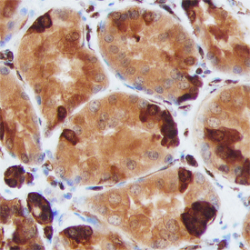
S100A6 in Human Kidney. S100A6 was detected in immersion fixed paraffin-embedded sections of human kidney using Sheep Anti-Human/Mouse S100A6 Antigen Affinity-purified Polyclonal Antibody (Catalog # AF4584) at 3 µg/mL overnight at 4 °C. Before incubation with the primary antibody, tissue was subjected to heat-induced epitope retrieval using Antigen Retrieval Reagent-Basic (Catalog # CTS013). Tissue was stained using the Anti-Sheep HRP-DAB Cell & Tissue Staining Kit (brown; Catalog # CTS019) and counterstained with hematoxylin (blue). Specific staining was localized to epithelial cells in convoluted tubules. View our protocol for Chromogenic IHC Staining of Paraffin-embedded Tissue Sections.
 View Larger
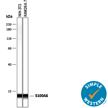
Detection of Mouse S100A6 by Simple WesternTM. Simple Western lane view shows lysates of NIH-3T3 mouse embryonic fibroblast cell line and RAW 264.7 mouse monocyte/macrophage cell line, loaded at 0.2 mg/mL. A specific band was detected for S100A6 at approximately 7 kDa (as indicated) using 10 µg/mL of Sheep Anti-Human/Mouse S100A6 Antigen Affinity-purified Polyclonal Antibody (Catalog # AF4584) followed by 1:50 dilution of HRP-conjugated Anti-Sheep IgG Secondary Antibody (Catalog # HAF016). This experiment was conducted under reducing conditions and using the 12-230 kDa separation system.
 View Larger
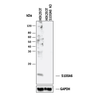
Western Blot Shows Human S100A6 Specificity by Using Knockout Cell Line. Western blot shows lysates of HEK293T human embryonic kidney parental cell line and S100A6 knockout HEK293T cell line (KO). PVDF membrane was probed with 1 µg/mL of Sheep Anti-Human/Mouse S100A6 Antigen Affinity-purified Polyclonal Antibody (Catalog # AF4584) followed by HRP-conjugated Anti-Sheep IgG Secondary Antibody (Catalog # HAF016). A specific band was detected for S100A6 at approximately 10 kDa (as indicated) in the parental HEK293T cell line, but is not detectable in knockout HEK293T cell line. GAPDH (Catalog # AF5718) is shown as a loading control. This experiment was conducted under reducing conditions and using Immunoblot Buffer Group 1.
Reconstitution Calculator
Preparation and Storage
- 12 months from date of receipt, -20 to -70 degreesC as supplied. 1 month, 2 to 8 degreesC under sterile conditions after reconstitution. 6 months, -20 to -70 degreesC under sterile conditions after reconstitution.
Background: S100A6
Mouse S100A6 (also prolactin receptor-associated protein and calcyclin) is a 10 kDa member of the S100 family of calcium-binding proteins. S100A6 is 89 amino acids (aa) in length and contains two calcium-binding EF-hand domains (aa 12-47 and 48-83). Intracellularly, S100A6 will both noncovalently homodimerize and heterodimerize with S100B plus SGT1. Extracellularly, it is secreted via a noncanonical pathway and binds to RAGE, inducing apoptosis. It is expressed by neurons, endothelium, fibroblasts and glandular epithelia. Mouse S100A6 is 99% and 96% aa identical to rat and human S100A6, respectively.
Product Datasheets
Citations for Human/Mouse S100A6 Antibody
R&D Systems personnel manually curate a database that contains references using R&D Systems products. The data collected includes not only links to publications in PubMed, but also provides information about sample types, species, and experimental conditions.
2
Citations: Showing 1 - 2
Filter your results:
Filter by:
-
Circulating Ligands of the Receptor for Advanced Glycation End Products and the Soluble Form of the Receptor Modulate Cardiovascular Cell Apoptosis in Diabetes
Authors: JN Tsoporis, E Hatziagela, S Gupta, S Izhar, V Salpeas, A Tsiavou, AG Rigopoulos, AS Triantafyl, JC Marshall, TG Parker, IK Rizos
Molecules, 2020-11-10;25(22):.
Species: Human
Sample Types: Plasma
Applications: Plasma Culture -
MMP-8 Deficiency Increases TLR/RAGE Ligands S100A8 and S100A9 and Exacerbates Lung Inflammation during Endotoxemia.
Authors: Gonzalez-Lopez A, Aguirre A, Lopez-Alonso I, Amado L, Astudillo A, Fernandez-Garcia MS, Suarez MF, Batalla-Solis E, Colado E, Albaiceta GM
PLoS ONE, 2012-06-29;7(6):e39940.
Species: Mouse
Sample Types: Tissue Homogenates
Applications: Western Blot
FAQs
No product specific FAQs exist for this product, however you may
View all Antibody FAQsReviews for Human/Mouse S100A6 Antibody
There are currently no reviews for this product. Be the first to review Human/Mouse S100A6 Antibody and earn rewards!
Have you used Human/Mouse S100A6 Antibody?
Submit a review and receive an Amazon gift card.
$25/€18/£15/$25CAN/¥75 Yuan/¥2500 Yen for a review with an image
$10/€7/£6/$10 CAD/¥70 Yuan/¥1110 Yen for a review without an image

