Human MyD88 Antibody Summary
Met1-Pro296
Accession # Q99836
Applications
Please Note: Optimal dilutions should be determined by each laboratory for each application. General Protocols are available in the Technical Information section on our website.
Scientific Data
 View Larger
Detection of Human MyD88 by Western Blot. Western blot shows lysates of Raji human Burkitt's lymphoma cell line, Jurkat human acute T cell leukemia cell line, and HT-29 human colon adenocarcinoma cell line. PVDF membrane was probed with 0.5 µg/mL Goat Anti-Human MyD88 Antigen Affinity-purified Polyclonal Antibody (Catalog # AF2928) followed by HRP-conjugated Anti-Goat IgG Secondary Antibody (Catalog # HAF017). For additional reference, recombinant human MyD88 (1 ng) was included. A specific band for MyD88 was detected at approximately 35 kDa (as indicated). This experiment was conducted under reducing conditions and using Immunoblot Buffer Group 3.
.") View Larger
View Larger
MyD88 in Raji Human Cell Line. MyD88 was detected in immersion fixed Raji human Burkitt's lymphoma cell line using Goat Anti-Human MyD88 Antigen Affinity-purified Polyclonal Antibody (Catalog # AF2928) at 15 µg/mL for 3 hours at room temperature. Cells were stained using the NorthernLights™ 557-conjugated Anti-Goat IgG Secondary Antibody (red; Catalog # NL001) and counterstained with DAPI (blue). Specific staining was localized to cytoplasm. View our protocol for Fluorescent ICC Staining of Non-adherent Cells.
 View Larger
Detection of MyD88 in Raji Human Cell Line by Flow Cytometry. Raji human Burkitt's lymphoma cell line was stained with Goat Anti-Human MyD88 Antigen Affinity-purified Polyclonal Antibody (Catalog # AF2928, filled histogram) or isotype control antibody (Catalog # AB-108-C, open histogram), followed by Allophycocyanin-conjugated Anti-Goat IgG Secondary Antibody (Catalog # F0108). To facilitate intracellular staining, cells were fixed with paraformaldehyde and permeabilized with saponin.
 View Larger
Detection of Human MyD88 by Simple WesternTM. Simple Western lane view shows lysates of Raji human Burkitt's lymphoma cell line, loaded at 0.2 mg/mL. A specific band was detected for MyD88 at approximately 41 kDa (as indicated) using 5 µg/mL of Goat Anti-Human MyD88 Antigen Affinity-purified Polyclonal Antibody (Catalog # AF2928) followed by 1:50 dilution of HRP-conjugated Anti-Goat IgG Secondary Antibody (Catalog # HAF109). This experiment was conducted under reducing conditions and using the 12-230 kDa separation system.
 View Larger
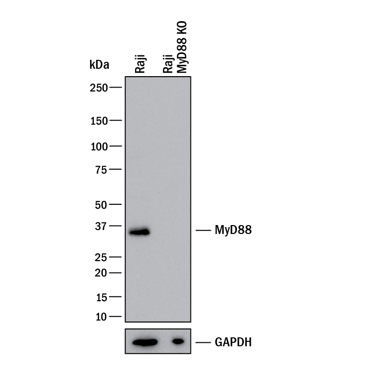
Western Blot Shows Human MyD88 Specificity by Using Knockout Cell Line. Western blot shows lysates of Raji human Burkitt's lymphoma cell line and human MyD88 knockout Raji human Burkitt's lymphoma cell line (KO). PVDF membrane was probed with 0.5 µg/mL of Goat Anti-Human MyD88 Antigen Affinity-purified Polyclonal Antibody (Catalog # AF2928) followed by HRP-conjugated Anti-Goat IgG Secondary Antibody (HAF017). A specific band was detected for MyD88 at approximately 35 kDa (as indicated) in the parental Raji human Burkitt's lymphoma cell line, but is not detectable in knockout Raji human Burkitt's lymphoma cell line. GAPDH (AF5718) is shown as a loading control. This experiment was conducted under reducing conditions and using Western Blot Buffer Group 1.
Reconstitution Calculator
Preparation and Storage
- 12 months from date of receipt, -20 to -70 °C as supplied.
- 1 month, 2 to 8 °C under sterile conditions after reconstitution.
- 6 months, -20 to -70 °C under sterile conditions after reconstitution.
Background: MyD88
Myeloid Differentiation primary response gene 88 (MyD88) is a 35 kDa adaptor protein involved in the IL-1 signaling pathway and recruits IRAK-2 to the IL-1 receptor complex. MyD88 also recruits IRAK-4 to Toll-like receptors 2 and 4 (TLR2 and TLR4) and plays an important role in the inflammatory response induced by IL-1, IL-18 and endotoxin.
Product Datasheets
Citations for Human MyD88 Antibody
R&D Systems personnel manually curate a database that contains references using R&D Systems products. The data collected includes not only links to publications in PubMed, but also provides information about sample types, species, and experimental conditions.
3
Citations: Showing 1 - 3
Filter your results:
Filter by:
-
Cell-Based Screen Identifies Human Interferon-Stimulated Regulators of Listeria monocytogenes Infection
PLoS Pathog, 2016-12-21;12(12):e1006102.
Species: Human
Sample Types: Cell Lysates
Applications: Western Blot -
Enterovirus 68 3C protease cleaves TRIF to attenuate antiviral responses mediated by Toll-like receptor 3.
Authors: Xiang Z, Li L, Lei X, Zhou H, Zhou Z, He B, Wang J
J Virol, 2014-03-26;88(12):6650-9.
Species: Human
Sample Types: Cell Lysates
Applications: Western Blot -
Potentiation of flagellin responses in gut epithelial cells by interferon-gamma is associated with STAT-independent regulation of MyD88 expression.
Authors: Bannon C, Davies PJ, Collett A, Warhurst G
Biochem. J., 2009-09-14;423(1):119-28.
Species: Human
Sample Types: Cell Lysates
Applications: Western Blot
FAQs
No product specific FAQs exist for this product, however you may
View all Antibody FAQsReviews for Human MyD88 Antibody
Average Rating: 5 (Based on 1 Review)
Have you used Human MyD88 Antibody?
Submit a review and receive an Amazon gift card.
$25/€18/£15/$25CAN/¥75 Yuan/¥2500 Yen for a review with an image
$10/€7/£6/$10 CAD/¥70 Yuan/¥1110 Yen for a review without an image
Filter by:


