Human Myeloperoxidase/MPO Antibody Summary
Val279-Ser745
Accession # P05164
Applications
Please Note: Optimal dilutions should be determined by each laboratory for each application. General Protocols are available in the Technical Information section on our website.
Scientific Data
 View Larger
Detection of Human Myeloperoxidase/MPO by Western Blot. Western blot shows lysates of HL-60 human acute promyelocytic leukemia cell line and human neutrophil. PVDF membrane was probed with 1 µg/mL of Mouse Anti-Human Myeloperoxidase/MPO Monoclonal Antibody (Catalog # MAB3174) followed by HRP-conjugated Anti-Mouse IgG Secondary Antibody (Catalog # HAF007). A specific band was detected for Myeloperoxidase/MPO at approximately 65 kDa (as indicated). This experiment was conducted under reducing conditions and using Immunoblot Buffer Group 2.
 View Larger
Myeloperoxidase/MPO in MOLT-4 Human Cell Line. Myeloperoxidase/ MPO was detected in immersion fixed MOLT-4 human acute lymphoblastic leukemia cell line using 8 µg/mL Mouse Anti-Human Myeloperoxidase/ MPO Monoclonal Antibody (Catalog # MAB3174) for 3 hours at room temperature. Cells were stained (red) and counter-stained (green). View our protocol for Fluorescent ICC Staining of Cells on Coverslips.
 View Larger
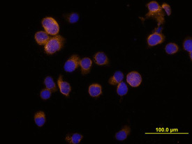
Myeloperoxidase/MPO in HL‑60 Human Cell Line. Myeloperoxidase/MPO was detected in immersion fixed HL-60 human acute promyelocytic leukemia cell line using Mouse Anti-Human Myeloperoxidase/MPO Mono-clonal Antibody (Catalog # MAB3174) at 10 µg/mL for 3 hours at room temperature. Cells were stained using the NorthernLights™ 557-conju-gated Anti-Mouse IgG Secondary Antibody (yellow; Catalog # NL007) and counterstained with DAPI (blue). View our protocol for Fluorescent ICC Staining of Non-adherent Cells.
 View Larger
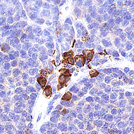
Myeloperoxidase/MPO in Human Spleen. Myeloperoxidase/MPO was detected in immersion fixed paraffin-embedded sections of human spleen using 15 µg/mL Mouse Anti-Human Myeloperoxidase/MPO Monoclonal Antibody (Catalog # MAB3174) overnight at 4 °C. Before incubation with the primary antibody tissue was subjected to heat-induced epitope retrieval using Antigen Retrieval Reagent-Basic (Catalog # CTS013). Tissue was stained with the Anti-Mouse HRP-DAB Cell & Tissue Staining Kit (brown; Catalog # CTS002) and counterstained with hematoxylin (blue). View our protocol for Chromogenic IHC Staining of Paraffin-embedded Tissue Sections.
 View Larger
Myeloperoxidase/MPO in Mouse Spleen. Myeloperoxidase/MPO was detected in immersion fixed paraffin-embedded sections of mouse spleen using Mouse Anti-Human Myeloperoxidase/MPO Monoclonal Antibody (Catalog # MAB3174) at 10 µg/mL for 1 hour at room temperature followed by incubation with the Anti-Mouse IgG VisUCyte™ HRP Polymer Antibody (VC001). Before incubation with the primary antibody, tissue was subjected to heat-induced epitope retrieval using Antigen Retrieval Reagent-Basic (CTS013). Tissue was stained using DAB (brown) and counterstained with hematoxylin (blue). Specific staining was localized to cytoplasm in lymphocytes. Staining was performed using our protocol for IHC Staining with VisUCyte HRP Polymer Detection Reagents.
Reconstitution Calculator
Preparation and Storage
- 12 months from date of receipt, -20 to -70 °C as supplied.
- 1 month, 2 to 8 °C under sterile conditions after reconstitution.
- 6 months, -20 to -70 °C under sterile conditions after reconstitution.
Background: Myeloperoxidase/MPO
Myeloperoxidase (MPO) is a hemeprotein that belongs to the XPO subfamily of the heme peroxidase superfamily. MPO is synthesized as a preproprotein that undergoes proteolytic processing to generate a disulfide-linked heterodimer of the N-terminal beta -subunit (12 kDa) and C-terminal alpha subunit (60 kDa). Active MPO is a tetramer of two beta -subunits and two alpha -subunits that are also disulfide-linked through the two alpha -subunits. MPO is stored in granules and is an abundant protein in neutrophils and monocytes. MPO is released upon activation to catalyze the formation of powerful oxidants such as hypochlorous acid, which kills microbes. Unprocessed pro-MPO can also be released. Human and mouse MPO share 87% amino acid sequence identity.
Product Datasheets
Citations for Human Myeloperoxidase/MPO Antibody
R&D Systems personnel manually curate a database that contains references using R&D Systems products. The data collected includes not only links to publications in PubMed, but also provides information about sample types, species, and experimental conditions.
10
Citations: Showing 1 - 10
Filter your results:
Filter by:
-
Shifting focus from bacteria to host neutrophil extracellular traps of biodegradable pure Zn to combat implant centered infection
Authors: F Peng, J Xie, H Liu, Y Zheng, X Qian, R Zhou, H Zhong, Y Zhang, M Li
Bioactive materials, 2022-09-15;21(0):436-449.
Species: Mouse
Sample Types: Cell Culture Supernates
Applications: ELISA Capture -
Iraq/Afghanistan war lung injury reflects burn pits exposure
Authors: T Olsen, D Caruana, K Cheslack-P, A Szema, J Thieme, A Kiss, M Singh, G Smith, S McClain, T Glotch, M Esposito, R Promisloff, D Ng, X He, M Egeblad, R Kew, A Szema
Scientific Reports, 2022-08-29;12(1):14671.
Species: Human
Sample Types: Whole Tissue
Applications: IHC -
Treatment of necrotizing enterocolitis by conditioned medium derived from human amniotic fluid stem cells
Authors: JS O'Connell, B Li, A Zito, A Ahmed, M Cadete, N Ganji, E Lau, M Alganabi, N Farhat, C Lee, S Eaton, R Mitchell, S Ray, P De Coppi, K Patel, A Pierro
PLoS ONE, 2021-12-02;16(12):e0260522.
Species: Human
Sample Types: Whole Tissue
Applications: IHC -
A novel histopathological scoring system to distinguish urticarial vasculitis from chronic spontaneous urticaria
Authors: V Puhl, H Bonnekoh, J Scheffel, T Hawro, K Weller, P von den Dr, HJ Röwert-Hub, J Cardoso, M Gonçalo, M Maurer, K Krause
Clinical and translational allergy, 2021-04-01;11(2):e12031.
Species: Human
Sample Types: Whole Tissue
Applications: IHC -
Cohort Analysis of ADAM8 Expression in the PDAC Tumor Stroma
Authors: C Jaworek, Y Verel-Yilm, S Driesch, S Ostgathe, L Cook, S Wagner, DK Bartsch, EP Slater, JW Bartsch
Journal of personalized medicine, 2021-02-10;11(2):.
Species: Human
Sample Types: Whole Tissue
Applications: IHC -
The Traditional Chinese Medicine MLC901 inhibits inflammation processes after focal cerebral ischemia
Authors: C Widmann, C Gandin, A Petit-Pait, M Lazdunski, C Heurteaux
Sci Rep, 2018-12-24;8(1):18062.
Species: Mouse
Sample Types: Whole Tissue
Applications: IHC -
Suppression Colitis and Colitis-Associated Colon Cancer by Anti-S100a9 Antibody in Mice
Authors: X Zhang, L Wei, J Wang, Z Qin, J Wang, Y Lu, X Zheng, Q Peng, Q Ye, F Ai, P Liu, S Wang, G Li, S Shen, J Ma
Front Immunol, 2017-12-13;8(0):1774.
Species: Mouse
Sample Types: Whole Tissue
Applications: IHC-P -
Antimicrobial cathelicidin peptide LL?37 induces NET formation and suppresses the inflammatory response in a mouse septic model
Authors: H Hosoda, K Nakamura, Z Hu, H Tamura, J Reich, K Kuwahara-A, T Iba, Y Tabe, I Nagaoaka
Mol Med Rep, 2017-08-17;0(0):.
Species: Human
Sample Types:
-
Myocardial Structural and Biological Anomalies Induced by High Fat Diet in Psammomys obesus Gerbils.
Authors: Sahraoui A, Dewachter C, de Medina G, Naeije R, Aouichat Bouguerra S, Dewachter L
PLoS ONE, 2016-02-03;11(2):e0148117.
Species: Gerbil
Sample Types: Whole Tissue
Applications: IHC-P -
The Ancient Immunoglobulin Domains of Peroxidasin Are Required to Form Sulfilimine Cross-links in Collagen IV.
Authors: Ero-Tolliver I, Hudson B, Bhave G
J Biol Chem, 2015-07-15;290(35):21741-8.
Species: Human
Sample Types: Cell Lysates
Applications: Western Blot
FAQs
No product specific FAQs exist for this product, however you may
View all Antibody FAQsReviews for Human Myeloperoxidase/MPO Antibody
Average Rating: 3.4 (Based on 5 Reviews)
Have you used Human Myeloperoxidase/MPO Antibody?
Submit a review and receive an Amazon gift card.
$25/€18/£15/$25CAN/¥75 Yuan/¥2500 Yen for a review with an image
$10/€7/£6/$10 CAD/¥70 Yuan/¥1110 Yen for a review without an image
Filter by:
This antibody does not stain Neutrophil Extracellular Traps (NETs) on coverslips, whereas it does stain intracellular MPO. This raises questions about its specificity. This antibody is also not reactive in a sandwich ELISA to detect NETs, despite the fact that MPO is known to be a very abundant protein on NETs... On western blot it does detect a protein at the right molecular weight already at low concentrations.
R&D Systems Technical Service is investigating.
Used at a dilution of 1:50. Non-R-D secondary, biotinylated at a dilution of 1:200
None


