Human NKp30/NCR3 Antibody Summary
Leu19-Thr138
Accession # Q05D23
Applications
Please Note: Optimal dilutions should be determined by each laboratory for each application. General Protocols are available in the Technical Information section on our website.
Scientific Data
 View Larger
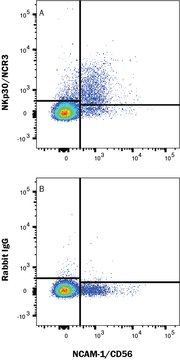
Detection of NKp30/NCR3 in Human PBMCs by Flow Cytometry. Human peripheral blood mononuclear cells (PBMCs) were stained with Mouse Anti-Human NCAM-1/CD56 PE-conjugated Monoclonal Antibody (Catalog # FAB2408P) and either (A) Rabbit Anti-Human NKp30/NCR3 Monoclonal Antibody (Catalog # MAB18492) or (B) Rabbit IgG Isotype Control (Catalog # MAB1050) followed by Goat anti-Rabbit IgG APC-conjugated secondary antibody (Catalog # F0111). View our protocol for Staining Membrane-associated Proteins.
 View Larger
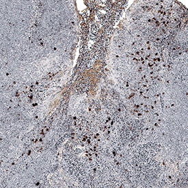
NKp30/NCR3 in Human Tonsil. NKp30/NCR3 was detected in immersion fixed paraffin-embedded sections of human tonsil using Rabbit Anti-Human NKp30/NCR3 Monoclonal Antibody (Catalog # MAB18492) at 1 µg/mL for 1 hour at room temperature followed by incubation with the Anti-Rabbit IgG VisUCyte™ HRP Polymer Antibody (Catalog # VC003). Before incubation with the primary antibody, tissue was subjected to heat-induced epitope retrieval using Antigen Retrieval Reagent-Basic (Catalog # CTS013). Tissue was stained using DAB (brown) and counterstained with hematoxylin (blue). Specific staining was localized to cell surface in lymphocytes. View our protocol for IHC Staining with VisUCyte HRP Polymer Detection Reagents.
Reconstitution Calculator
Preparation and Storage
- 12 months from date of receipt, -20 to -70 °C as supplied.
- 1 month, 2 to 8 °C under sterile conditions after reconstitution.
- 6 months, -20 to -70 °C under sterile conditions after reconstitution.
Background: NKp30/NCR3
NKp30, along with NKp44 and NKp46, constitute a group of receptors termed “Natural Cytotoxicity Receptors” (1). These receptors play a major role in triggering NK-mediated killing of most tumor cells lines. NKp30 is a type I transmembrane protein having a single extracellular V-like immunoglobulin domain (2). A physical association with the ITAM‑bearing accessory protein, CD3 zeta, occurs via a charged residue in the NKp30 transmembrane domain. Ligation of NKp30 with a specific antibody results in phosphorylation of CD3 zeta (3). NKp30 is expressed on both resting and activated NK cells of the CD56dim, CD16+ subset that account for more that 85% of NK cells found in peripheral blood and spleen (4). NKp30 is absent from the CD56bright, CD16- subset that constitutes the majority of NK cells in lymph node and tonsil, however, its expression is up-regulated in these cells upon IL-2 activation (4). Studies with neutralizing antibodies reveal that NKp30 is partially responsible for triggering lytic activity against several tumor cell types and that it is the main receptor responsible for NK-mediated lysis of immature dendritic cells (2, 5). The ligand(s) recognized by NKp30 has not been described.
- Moretta, L. and A. Moretta (2004) EMBO J. 23:255.
- Pende, D. et al. (1999) J. Exp. Med. 190:1505.
- Augugliaro, R. et al. (2003) Eur. J. Immunol. 33:1235.
- Ferlazzo, G. et al. (2004) J. Immunol. 172:1455.
- Ferlazzo, G. et al. (2002) J. Exp. Med. 195:343.
Product Datasheets
FAQs
No product specific FAQs exist for this product, however you may
View all Antibody FAQsReviews for Human NKp30/NCR3 Antibody
There are currently no reviews for this product. Be the first to review Human NKp30/NCR3 Antibody and earn rewards!
Have you used Human NKp30/NCR3 Antibody?
Submit a review and receive an Amazon gift card.
$25/€18/£15/$25CAN/¥75 Yuan/¥2500 Yen for a review with an image
$10/€7/£6/$10 CAD/¥70 Yuan/¥1110 Yen for a review without an image


