Human OSMR beta Antibody Summary
Glu28-Ser739
Accession # Q99650
Applications
Please Note: Optimal dilutions should be determined by each laboratory for each application. General Protocols are available in the Technical Information section on our website.
Scientific Data
 View Larger
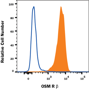
Detection of OSM R beta in HeLa Human Cell Line by Flow Cytometry. HeLa human cervical epithelial carcinoma cell line was stained with Goat Anti-Human OSM R beta Antigen Affinity-purified Polyclonal Antibody (Catalog # AF4389, filled histogram) or isotype control antibody (Catalog # AB-108-C, open histogram), followed by Phycoerythrin-conjugated Anti-Goat IgG Secondary Antibody (Catalog # F0107).
 View Larger
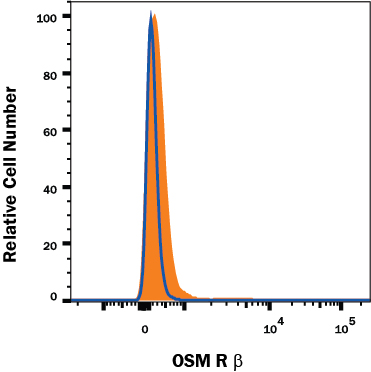
OSM R beta Specificity is Shown by Flow Cytometry in Knockout Cell Line. OSM R beta knockout HeLa epithelial carcinoma cell line was stained with Mouse Anti-Human OSM R beta Affinity Purified Polyclonal Antibody (Catalog # AF4389, filled histogram) or isotype control antibody (Catalog # AB-108-C, open histogram) followed by PE-conjugated anti-Goat IgG Secondary Antibody (Catalog # F0107). No staining in the OSM R beta knockout HeLa cell line was observed. View our protocol for Staining Membrane-associated Proteins.
Reconstitution Calculator
Preparation and Storage
- 12 months from date of receipt, -20 to -70 °C as supplied.
- 1 month, 2 to 8 °C under sterile conditions after reconstitution.
- 6 months, -20 to -70 °C under sterile conditions after reconstitution.
Background: OSMR beta
OSM R beta is a 150‑180 kDa member of the IL-6 receptor family. It associates with gp130 to form the type II OSM receptor that is responsive to OSM. The gp130 subunit is shared by other IL-6 family cytokine receptors (1, 2, 3, 4), and OSM R beta associates with gp130-like receptor (GPL) to form a receptor complex responsive to IL-31 (5, 6). The human OSM R beta cDNA encodes a 979 amino acid (aa) precursor that includes a 27 aa signal sequence, a 712 aa extracellular domain (ECD), a 22 aa transmembrane segment, and a 218 aa cytoplasmic domain. The ECD contains one partial and one complete hematopoietin domain, an Ig-like domain, and three fibronectin type-III domains. The cytoplasmic domain contains box1, 2, and 3 motifs (7). Within the ECD, human OSM R beta shares 55%, 58%, 61%, and 72% aa sequence identity with mouse, rat, bovine, and canine OSM R beta, respectively. It also shares 31% aa sequence identity with human LIF R, but less than 20% aa sequence identity with human CNTF R alpha, G-CSF R, IL-6 R, IL-11 R alpha, and TCCR. OSM R beta does not bind cytokines directly, but increases the affinity of gp130 for OSM, and GPL for IL-31 (7, 8). OSM R beta, gp130, and GPL each initiate signaling events following ligand stimulation (9, 10). Jak/STAT and MAPK pathways are activated by OSM R beta -containing receptors (9, 11, 12, 13), including STAT5b and SHC which are not activated by other IL-6 family receptors (10, 13). In mice, the loss of OSM R beta expression blocks erythroid progenitor development in bone marrow, and dramatically reduces the number of circulating platelets and erythrocytes (14). The type II OSM receptor is the only IL-6 family receptor that promotes osteoblast differentiation in calvaria cell cultures (15).
- Chen, S.-H. and E.N. Benveniste (2004) Cytokine Growth Factor Rev. 15:379.
- Heinrich, P.C. et al. (2003) Biochem. J. 374:1.
- Tanaka, M. and A. Miyajima (2003) Rev. Physiol. Biochem. Pharmacol. 149:39.
- Gearing, D.P. et al. (1992) Science 255:1434.
- Dillon, S.R. et al. (2004) Nat. Immunol. 5:752.
- Diveu, C. et al. (2003) J. Biol. Chem. 278:49850.
- Mosley, B. et al. (1996) J. Biol. Chem. 271:32635.
- Diveu, C. et al. (2004) Eur. Cytokine Netw. 15:291.
- Dreuw, A. et al. (2004) J. Biol. Chem. 279:36112.
- Wang, Y. et al. (2000) J. Biol. Chem. 275:25273.
- Hermanns, H.M. et al. (2000) J. Biol. Chem. 275:40742.
- Kuropatwinski, K.K. et al. (1997) J. Biol. Chem. 272:15135.
- Auguste, P. et al. (1997) J. Biol. Chem. 272:15760.
- Tanaka, M. et al. (2003) Blood 102:3154.
- Malaval, L. et al. (2005) J. Cell. Physiol. 204:585.
Product Datasheets
FAQs
No product specific FAQs exist for this product, however you may
View all Antibody FAQsReviews for Human OSMR beta Antibody
Average Rating: 2 (Based on 1 Review)
Have you used Human OSMR beta Antibody?
Submit a review and receive an Amazon gift card.
$25/€18/£15/$25CAN/¥75 Yuan/¥2500 Yen for a review with an image
$10/€7/£6/$10 CAD/¥70 Yuan/¥1110 Yen for a review without an image
Filter by:
Tried with various conditions on cells lysates without success by Western blot. However, when I tried an OSMR antibody from a different company on the same samples alongside, that worked no problem. The antibody was only tried for Western blot.


