Human Osterix/Sp7 Antibody Summary
Met19-Leu288
Accession # Q8TDD2
Applications
Please Note: Optimal dilutions should be determined by each laboratory for each application. General Protocols are available in the Technical Information section on our website.
Scientific Data
 View Larger
Detection of Human Osterix/Sp7 by Western Blot. Western blot shows lysates of Saos-2 human osteosarcoma cell line. PVDF membrane was probed with 1 µg/mL of Mouse Anti-Human Osterix/Sp7 Monoclonal Antibody (Catalog # MAB7547) followed by HRP-conjugated Anti-Mouse IgG Secondary Antibody (Catalog # HAF018). A specific band was detected for Osterix/Sp7 at approximately 45 kDa (as indicated). This experiment was conducted under reducing conditions and using Immunoblot Buffer Group 1.
 View Larger
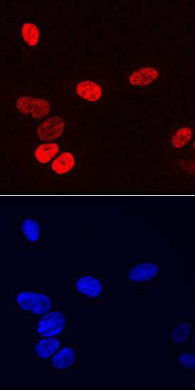
Osterix/Sp7 in Saos‑2 Human Cell Line. Osterix/Sp7 was detected in immersion fixed Saos-2 human osteosarcoma cell line using Mouse Anti-Human Osterix/Sp7 Monoclonal Antibody (Catalog # MAB7547) at 10 µg/mL for 3 hours at room temperature. Cells were stained using the NorthernLights™ 557-conjugated Anti-Mouse IgG Secondary Antibody (red, upper panel; Catalog # NL007) and counterstained with DAPI (blue, lower panel). Specific staining was localized to nuclei. View our protocol for Fluorescent ICC Staining of Cells on Coverslips.
Reconstitution Calculator
Preparation and Storage
- 12 months from date of receipt, -20 to -70 °C as supplied.
- 1 month, 2 to 8 °C under sterile conditions after reconstitution.
- 6 months, -20 to -70 °C under sterile conditions after reconstitution.
Background: Osterix/Sp7
Osterix, also known as Sp7, is an approximately 45 kDa transcription factor that is required for osteogenesis and bone homeostasis. Osterix cooperates with BMP-6 in the differentiation of osteoblasts from mesenchymal stem cells by regulating the transcription of several genes involved in osteoblast differentiation and function (i.e. SATB2, Collagen V, and SOST). The transcription of Osterix is induced by BMP-2, IGF-I, and parathyroid hormone, while its activity is regulated by Akt and p38 MAPK-mediated phoshorylation. Osterix contains a transactivation domain (aa 141-210) and three zinc finger domains (aa 294-318, aa 324-348, and aa 354-376). Alternative splicing generates a short isoform that lacks the N-terminal 18 amino acids. Within aa 19-288, human Osterix shares 94% aa sequence identity with mouse and rat Osterix.
Product Datasheets
Citation for Human Osterix/Sp7 Antibody
R&D Systems personnel manually curate a database that contains references using R&D Systems products. The data collected includes not only links to publications in PubMed, but also provides information about sample types, species, and experimental conditions.
1 Citation: Showing 1 - 1
-
L-PRF Secretome from Both Smokers/Nonsmokers Stimulates Angiogenesis and Osteoblast Differentiation In Vitro
Authors: Ríos, S;González, LG;Saez, CG;Smith, PC;Escobar, LM;Martínez, CE;
Biomedicines
Species: Human
Sample Types: Cell Lysates, Whole Cells
Applications: Immunocytochemistry, Western Blot
FAQs
No product specific FAQs exist for this product, however you may
View all Antibody FAQsReviews for Human Osterix/Sp7 Antibody
There are currently no reviews for this product. Be the first to review Human Osterix/Sp7 Antibody and earn rewards!
Have you used Human Osterix/Sp7 Antibody?
Submit a review and receive an Amazon gift card.
$25/€18/£15/$25CAN/¥75 Yuan/¥2500 Yen for a review with an image
$10/€7/£6/$10 CAD/¥70 Yuan/¥1110 Yen for a review without an image


