Human/Mouse/Rat p70 S6 Kinase Antibody Summary
Applications
Please Note: Optimal dilutions should be determined by each laboratory for each application. General Protocols are available in the Technical Information section on our website.
Scientific Data
 View Larger
Detection of Human and Mouse p70 S6 Kinase by Western Blot. Western blot shows lysates of MCF-7 human breast cancer cell line, HeLa human cervical epithelial carcinoma cell line, and NIH-3T3 mouse embryonic fibroblast cell line. PVDF membrane was probed with 0.2 µg/mL of Rabbit Anti-Human/Mouse/Rat p70 S6 Kinase Antigen Affinity-purified Polyclonal Antibody (Catalog # AF8962) followed by HRP-conjugated Anti-Rabbit IgG Secondary Antibody (Catalog # HAF008). A specific band was detected for p70 S6 Kinase at approximately 70 and 90 kDa (as indicated). This experiment was conducted under reducing conditions and using Immunoblot Buffer Group 1.
 View Larger
Detection of p70 S6 Kinase in HeLa Human Cell Line by Flow Cytometry. HeLa human cervical epithelial carcinoma cell line was stained with Rabbit Anti-Human/Mouse/Rat p70 S6 Kinase Antigen Affinity-purified Polyclonal Antibody (Catalog # AF8962, filled histogram) or control antibody (Catalog # AB-105-C, open histogram), followed by Phycoerythrin-conjugated Anti-Rabbit IgG Secondary Antibody (Catalog # F0110). To facilitate intracellular staining, cells were fixed with paraformaldehyde and permeabilized with methanol.
 View Larger
p70 S6 Kinase in Human Breast Cancer Tissue. p70 S6 Kinase was detected in immersion fixed paraffin-embedded sections of human breast cancer tissue using Rabbit Anti-Human/Mouse/Rat p70 S6 Kinase Antigen Affinity-purified Polyclonal Antibody (Catalog # AF8962) at 1.7 µg/mL overnight at 4 °C. Tissue was stained using the Anti-Rabbit HRP-DAB Cell & Tissue Staining Kit (brown; Catalog # CTS005) and counterstained with hematoxylin (blue). Specific labeling was localized to the nuclei of epithelial cells. View our protocol for Chromogenic IHC Staining of Paraffin-embedded Tissue Sections.
 View Larger
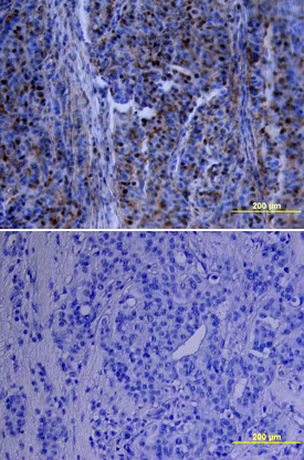
p70 S6 Kinase in Human Breast Cancer Tissue. p70 S6 Kinase was detected in immersion fixed paraffin-embedded sections of human breast cancer tissue using Rabbit Anti-Human/Mouse/Rat p70 S6 Kinase Antigen Affinity-purified Polyclonal Antibody (Catalog # AF8962) at 15 µg/mL overnight at 4 °C. Tissue was stained using the Anti-Rabbit HRP-DAB Cell & Tissue Staining Kit (brown; Catalog # CTS005) and counterstained with hematoxylin (blue). Lower panel shows a lack of labeling if primary antibodies are omitted and tissue is stained only with secondary antibody followed by incubation with detection reagents. View our protocol for Chromogenic IHC Staining of Paraffin-embedded Tissue Sections.
 View Larger
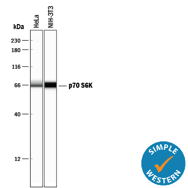
Detection of p70 S6 Kinase by Simple WesternTM. Simple Western lane view shows lysates of HeLa human cervical epithelial carcinoma cell line and NIH‑3T3 mouse embryonic fibroblast cell line, loaded at 0.2 mg/mL. A specific band was detected for p70 S6 Kinase at approximately 66 kDa (as indicated) using 2 µg/mL of Rabbit Anti-Human/Mouse/Rat p70 S6 Kinase Antigen Affinity-purified Polyclonal Antibody (Catalog # AF8962). This experiment was conducted under reducing conditions and using the 12-230 kDa separation system.
Reconstitution Calculator
Preparation and Storage
- 12 months from date of receipt, -20 to -70 degreesC as supplied. 1 month, 2 to 8 degreesC under sterile conditions after reconstitution. 6 months, -20 to -70 degreesC under sterile conditions after reconstitution.
Background: p70 S6 Kinase
p70 S6 kinase is a Ser/Thr kinase activated by such mitogens as EGF, IGF-I, and insulin. The S6 protein of the 40S ribosomal subunit is a major substrate of p70 S6 kinase, and its phosphorylation upregulates the translation of mRNAs containing an oligopyrimidine tract at their transcriptional start site. The activity of p70 S6 kinase is controlled by multiple phosphorylation events, with phosphorylation at T229 by PDK1 and T389 by mTOR most critical for kinase function.
Product Datasheets
Citations for Human/Mouse/Rat p70 S6 Kinase Antibody
R&D Systems personnel manually curate a database that contains references using R&D Systems products. The data collected includes not only links to publications in PubMed, but also provides information about sample types, species, and experimental conditions.
4
Citations: Showing 1 - 4
Filter your results:
Filter by:
-
Characterizing the distributions of IDO-1 expressing macrophages/microglia in human and murine brains and evaluating the immunological and physiological roles of IDO-1 in RAW264.7/BV-2 cells
Authors: R Ji, L Ma, X Chen, R Sun, L Zhang, H Saiyin, W Wei
PLoS ONE, 2021-11-04;16(11):e0258204.
Species: Human
Sample Types: Cell Lysates
Applications: Western Blot -
Trehalose limits opportunistic mycobacterial survival during HIV co-infection by reversing HIV-mediated autophagy block
Authors: V Sharma, M Makhdoomi, L Singh, P Kumar, N Khan, S Singh, HN Verma, K Luthra, S Sarkar, D Kumar
Autophagy, 2020-02-20;0(0):1-20.
Species: Human
Sample Types:
-
Mild MPP(+) exposure-induced glucose starvation enhances autophagosome synthesis and impairs its degradation
Authors: S Sakamoto, M Miyara, S Sanoh, S Ohta, Y Kotake
Sci Rep, 2017-04-26;7(0):46668.
Species: Human
Sample Types: Cell Lysates
Applications: Western Blot -
The role of autophagy in cardiomyocytes in the basal state and in response to hemodynamic stress.
Authors: Nakai A, Yamaguchi O, Takeda T
Nat. Med., 2007-04-22;13(5):619-24.
Species: Mouse
Sample Types: Cell Lysates
Applications: Western Blot
FAQs
No product specific FAQs exist for this product, however you may
View all Antibody FAQsReviews for Human/Mouse/Rat p70 S6 Kinase Antibody
Average Rating: 5 (Based on 3 Reviews)
Have you used Human/Mouse/Rat p70 S6 Kinase Antibody?
Submit a review and receive an Amazon gift card.
$25/€18/£15/$25CAN/¥75 Yuan/¥2500 Yen for a review with an image
$10/€7/£6/$10 CAD/¥70 Yuan/¥1110 Yen for a review without an image
Filter by:


