Human PAK1 Antibody Summary
Leu128-Gln242
Accession # Q13153
Applications
Please Note: Optimal dilutions should be determined by each laboratory for each application. General Protocols are available in the Technical Information section on our website.
Scientific Data
 View Larger
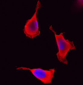
PAK1 in HeLa Human Cell Line. PAK1 was detected in immersion fixed HeLa human cervical epithelial carcinoma cell line using Goat Anti-Human PAK1 Antigen Affinity-purified Polyclonal Antibody (Catalog # AF7495) at 15 µg/mL for 3 hours at room temperature. Cells were stained using the NorthernLights™ 557-conjugated Anti-Goat IgG Secondary Antibody (red; Catalog # NL001) and counterstained with DAPI (blue). Specific staining was localized to cytoplasm. View our protocol for Fluorescent ICC Staining of Cells on Coverslips.
Reconstitution Calculator
Preparation and Storage
- 12 months from date of receipt, -20 to -70 °C as supplied.
- 1 month, 2 to 8 °C under sterile conditions after reconstitution.
- 6 months, -20 to -70 °C under sterile conditions after reconstitution.
Background: PAK1
PAK1 (p21-activaed kinase 1; also alpha -PAK and p65-PAK) is both a cytoplasmic and nuclear 58-60 kDa member of PAK group I, STE20 subfamily, STE Ser/Thr protein kinase family of molecules. It is widely expressed, and is upregulated in numerous cancers. PAK1 has multiple activities. It is responsible for regulating focal adhesions during motility. At the front edge of cells, PAK1 promotes focal adhesion formation by phosphorylating PIX, while at the back edge, it directs focal adhesion disassembly. PAK1 also impacts microtubule formation. Microtubules are composed of heterodimeric subunits containing an alpha - and beta -tubulin polypeptide. The availability of alpha -tubulin for microtubule formation is based on the ability of PAK1 to phosphorylate a alpha -tubulin chaperone, tubulin cofactor B. PAK1 is activated following binding to active GTPases which induce PAK autophosphorylation. When active, PAK1 is a monomer; when inactive, it reportedly homodimerizes. Human PAK1 is 545 amino acids (aa) in length. It contains one GTPase binding domain (aa 75-105) plus a protein kinase catalytic region (aa 250-521). There are five SH3-binding motifs. Acetylation occurs on Lys256, and there are at least 14 potential phosphorylation sites, seven of which are known to be utilized. There is one splice variant that shows a 35 aa substitution for aa 518-545. Over aa 128-242, human PAK1 shares 96% aa sequence identity with mouse PAK1.
Product Datasheets
FAQs
No product specific FAQs exist for this product, however you may
View all Antibody FAQsReviews for Human PAK1 Antibody
There are currently no reviews for this product. Be the first to review Human PAK1 Antibody and earn rewards!
Have you used Human PAK1 Antibody?
Submit a review and receive an Amazon gift card.
$25/€18/£15/$25CAN/¥75 Yuan/¥2500 Yen for a review with an image
$10/€7/£6/$10 CAD/¥70 Yuan/¥1110 Yen for a review without an image


