Human Pax6 Antibody Summary
Met1-Arg272
Accession # P26367
Applications
Please Note: Optimal dilutions should be determined by each laboratory for each application. General Protocols are available in the Technical Information section on our website.
Scientific Data
 View Larger
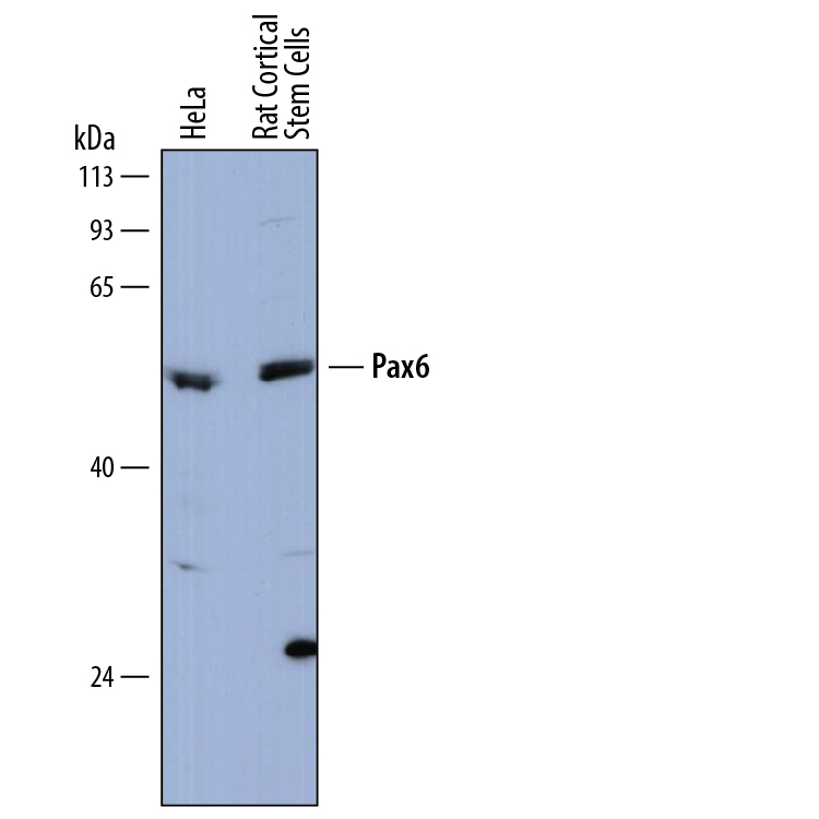
Detection of Human and Rat Pax6 by Western Blot. Western blot shows lysates of HeLa human cervical epithelial carcinoma cell line and rat cortical stem cells. PVDF membrane was probed with 0.5 µg/mL of Sheep Anti-Human Pax6 Antigen Affinity-purified Polyclonal Antibody (Catalog # AF8150) followed by HRP-conjugated Anti-Sheep IgG Secondary Antibody (Catalog # HAF016). A specific band was detected for Pax6 at approximately 48-50 kDa (as indicated). This experiment was conducted under reducing conditions and using Immunoblot Buffer Group 1.
 View Larger
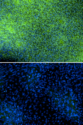
Pax6 in SA01 Human Embryonic Stem Cells. Immersion fixed SA01 human embryonic stem cells were differentiated for 6 days with Recombinant Human Noggin (Catalog # 6057-NG) and SB431542 (upper panel) or differentiated for 6 days with Recombinant Human BMP-4 (negative control, lower panel; Catalog # 314-BP). Pax6 was detected using Sheep Anti-Human Pax6 Antigen Affinity-purified Polyclonal Antibody (Catalog # AF8150) at 5 µg/mL. Cells were stained using an Alexa Fluor 488-conjugated Anti-Sheep IgG Secondary Antibody (green) and counterstained with DAPI (blue). Specific staining was localized to nuclei.Images courtesy of Dr. Ron McKay, Leiber Institute for Brain Development, Baltimore, Maryland, USA.
 View Larger
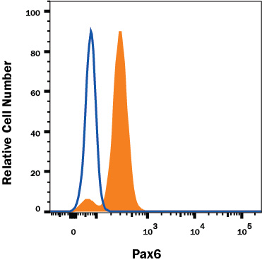
Detection of Pax6 in HeLa Human Cell Line by Flow Cytometry. HeLa human cervical epithelial carcinoma cell line was stained with Sheep Anti-Human Pax6 Affinity-Purified Polyclonal Antibody (Catalog # AF8150, filled histogram) or Sheep IgG control Antibody (Catalog # 5-001-A, open histogram) followed by anti-Sheep IgG PE-conjugated Secondary Antibody (Catalog # F0126). To facilitate intracellular staining, cells were fixed and permeabilized with FlowX FoxP3 Fixation & Permeabilization Buffer Kit (Catalog # FC012). View our protocol for Staining Intracellular Molecules.
 View Larger
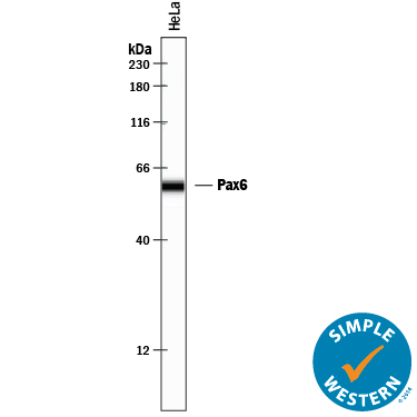
Detection of Human Pax6 by Simple WesternTM. Simple Western lane view shows lysates of HeLa human cervical epithelial carcinoma cell line, loaded at 0.2 mg/mL. A specific band was detected for Pax6 at approximately 59 kDa (as indicated) using 5 µg/mL of Sheep Anti-Human Pax6 Antigen Affinity-purified Polyclonal Antibody (Catalog # AF8150) followed by 1:50 dilution of HRP-conjugated Anti-Sheep IgG Secondary Antibody (Catalog # HAF016). This experiment was conducted under reducing conditions and using the 12-230 kDa separation system.
Reconstitution Calculator
Preparation and Storage
- 12 months from date of receipt, -20 to -70 °C as supplied.
- 1 month, 2 to 8 °C under sterile conditions after reconstitution.
- 6 months, -20 to -70 °C under sterile conditions after reconstitution.
Background: Pax6
Pax6 (paired box 6; also Oculorhombin) is a 48‑50 kDa member of the paired homeobox family of transcription factors. It is expressed in developing optic vesicle, olfactory dopaminergic neurons, and pancreatic endocrine cells. Pax6 is a transactivating protein that interacts with MAF, CDX2 and SOX2. Human Pax6 is 422 amino acids (aa) in length. It contains an N-terminal paired box DNA-binding domain (aa 4‑130), a Gly-rich central region (aa 131‑209), a homeodomain (aa 213‑269) and a C-terminal Pro/Ser/Thr-rich regulatory domain (aa 279‑422). Phosphorylation of the C‑terminal domain at Thr281/304/373 promotes Pax6 activity. Multiple splice forms of Pax6 exist. There are alternative start sites at Met137 and a position 34 aa upstream of the standard site. There is also a deletion of aa 22‑26 and 37‑39, plus a 14 aa insertion after Gln47 that generates a C‑terminal DNA binding site. Human and mouse Pax6 are absolutely identical in aa sequence.
Product Datasheets
Citations for Human Pax6 Antibody
R&D Systems personnel manually curate a database that contains references using R&D Systems products. The data collected includes not only links to publications in PubMed, but also provides information about sample types, species, and experimental conditions.
10
Citations: Showing 1 - 10
Filter your results:
Filter by:
-
Metabolic lactate production coordinates vasculature development and progenitor behavior in the developing mouse neocortex
Authors: X Dong, Q Zhang, X Yu, D Wang, J Ma, J Ma, SH Shi
Nature Neuroscience, 2022-06-20;0(0):.
Species: Mouse
Sample Types: Whole Tissue
Applications: IHC -
Down-syndrome-induced senescence disrupts the nuclear architecture of neural progenitors
Authors: HS Meharena, A Marco, V Dileep, ER Lockshin, GY Akatsu, J Mullahoo, LA Watson, T Ko, LN Guerin, F Abdurrob, S Rengarajan, M Papanastas, JD Jaffe, LH Tsai
Cell Stem Cell, 2022-01-06;29(1):116-130.e7.
Species: Human
Sample Types: Whole Cells
Applications: ICC -
Regulation of prefrontal patterning and connectivity by retinoic acid
Authors: M Shibata, K Pattabiram, B Lorente-Ga, D Andrijevic, SK Kim, N Kaur, SK Muchnik, X Xing, G Santpere, AMM Sousa, N Sestan
Nature, 2021-10-01;0(0):.
Species: Human
Sample Types: Whole Tissue
Applications: IHC -
SARS-CoV-2 infects human adult donor eyes and hESC-derived ocular epithelium
Authors: AZ Eriksen, R Møller, B Makovoz, SA Uhl, BR tenOever, TA Blenkinsop
Cell Stem Cell, 2021-05-17;0(0):.
Species: Human
Sample Types: Whole Tissue
Applications: IHC -
Organoid modeling of Zika and herpes simplex virus 1 infections reveals virus-specific responses leading to microcephaly
Authors: V Krenn, C Bosone, TR Burkard, J Spanier, U Kalinke, A Calistri, C Salata, R Rilo Chris, P Pestana Ga, A Mirazimi, JA Knoblich
Cell Stem Cell, 2021-04-09;0(0):.
Species: Human
Sample Types: Organoid
Applications: IHC -
Generation of 10 patient-specific induced pluripotent stem cells (iPSCs) to model Pitt-Hopkins Syndrome
Authors: SR Sripathy, Y Wang, RL Moses, A Fatemi, DA Batista, BJ Maher
Stem Cell Res, 2020-09-17;48(0):102001.
Species: Human
Sample Types: Whole Cells
Applications: ICC -
TALEN-mediated biallelic inactivation of MYB in human embryonic stem cell lines WAe001-A-45 and WAe001-A-46
Authors: C Fan, Z Shah, H Ullah, ES Philonenko, B Zhang, Y Tan, C Wang, J Zhang, IM Samokhvalo
Stem Cell Res, 2020-06-01;46(0):101854.
Species: Human
Sample Types: Whole Cells
Applications: ICC -
Chronic Exposure to Palmitate Impairs Insulin Signaling in an Intestinal L-cell Line: A Possible Shift from GLP-1 to Glucagon Production
Authors: A Filippello, F Urbano, S Di Mauro, A Scamporrin, A Di Pino, R Scicali, AM Rabuazzo, F Purrello, S Piro
Int J Mol Sci, 2018-11-28;19(12):.
Species: Mouse
Sample Types: Cell Lysates
Applications: Western Blot -
Generation of nine induced pluripotent stem cell lines as an ethnic diversity panel
Authors: X Gao, JJ Yourick, RL Sprando
Stem Cell Res, 2018-07-27;31(0):193-196.
Species: Human
Sample Types: Whole Cells
Applications: ICC -
TFAP2C regulates transcription in human naive pluripotency by opening enhancers
Authors: WA Pastor, W Liu, D Chen, J Ho, R Kim, TJ Hunt, A Lukianchik, X Liu, JM Polo, SE Jacobsen, AT Clark
Nat. Cell Biol., 2018-04-25;20(5):553-564.
Species: Human, Mouse
Sample Types: Cell Lysates, Whole Cells
Applications: IHC, Western Blot
FAQs
No product specific FAQs exist for this product, however you may
View all Antibody FAQsReviews for Human Pax6 Antibody
Average Rating: 4.3 (Based on 3 Reviews)
Have you used Human Pax6 Antibody?
Submit a review and receive an Amazon gift card.
$25/€18/£15/$25CAN/¥75 Yuan/¥2500 Yen for a review with an image
$10/€7/£6/$10 CAD/¥70 Yuan/¥1110 Yen for a review without an image
Filter by:
Pax6 was detected human embryonic stem cells-derived neural stem cells using Sheep Anti-Human PAX6 Antigen Affinity-purified Polyclonal Antibody (Catalog # AF8150) at 1 µg/mL for overnight at 4°C. Cells were stained using the Donkey anti-sheep IgG (H+L) Cross-Adsorbed, Alexa Fluor® 568.
Works fine at 1/1000 dilution. O.N. incubation at 4C.


