Human PD-L1 Antibody Summary
-H4, rhPD-L2, recombinant mouse B7-H1, recombinant rat (rr) B7-1, or rrB7-2 is observed.
Phe19-Thr239
Accession # Q9NZQ7
Applications
Please Note: Optimal dilutions should be determined by each laboratory for each application. General Protocols are available in the Technical Information section on our website.
Scientific Data
 View Larger
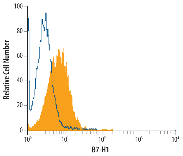
Detection of PD-L1/B7-H1 in Jurkat Human Cell Line by Flow Cytometry. Jurkat human acute T cell leukemia cell line was stained with Mouse Anti-Human PD-L1/B7-H1 Monoclonal Antibody (Catalog # MAB1561, filled histogram) or isotype control antibody (Catalog # MAB002, open histogram), followed by Phycoerythrin-conjugated Anti-Mouse IgG F(ab')2Secondary Antibody (Catalog # F0102B).View our protocol for Staining Membrane-associated Proteins.
 View Larger
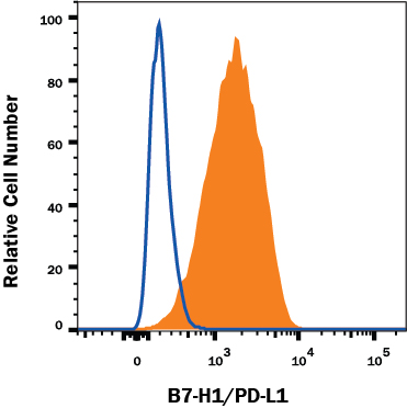
Detection of PD-L1/B7-H1 in MDA-MB-231 Human Cell Line by Flow Cytometry. MDA-MB-231 human breast adenocarcinoma cell line was stained with Mouse Anti-Human PD-L1/B7-H1 Monoclonal Antibody (Catalog # MAB1561, filled histogram) or isotype control antibody (Catalog # MAB002, open histogram), followed by Phycoerythrin-conjugated Anti-Mouse IgG F(ab')2 Secondary Antibody (Catalog # F0102B). Adherent cells were prepared by either manual scraping or with TrypLE Express treatment with similar results. View our protocol for Staining Membrane-associated Proteins.
 View Larger
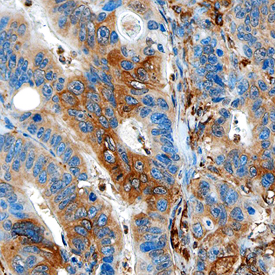
PD-L1/B7-H1 in Human Colon Cancer. PD-L1/B7-H1 was detected in formalin fixed paraffin-embedded sections of human colon cancer using Mouse Anti-Human PD-L1/B7-H1 Monoclonal Antibody (Catalog # MAB1561) at 15 µg/mL overnight at 4 °C. Tissue was stained using the Anti-Mouse HRP-DAB Cell & Tissue Staining Kit (brown; Catalog # CTS002) and counterstained with hematoxylin (blue). Specific staining was observed in the cytoplasm. View our protocol for Chromogenic IHC Staining of Paraffin-embedded Tissue Sections.
Reconstitution Calculator
Preparation and Storage
- 12 months from date of receipt, -20 to -70 °C as supplied.
- 1 month, 2 to 8 °C under sterile conditions after reconstitution.
- 6 months, -20 to -70 °C under sterile conditions after reconstitution.
Background: PD-L1/B7-H1
Human B7 homolog 1 (B7-H1), also called programmed death ligand 1 (PD-L1) and programmed cell death 1 ligand 1 (PDCD1L1), is a member of the growing B7 family of immune proteins that provide signals for both stimulating and inhibiting T cell activation. Other family members include B7-1, B7-2, B7-H2, PDL2 and B7-H3. B7 proteins are members of the immunoglobulin (Ig) superfamily. Their extracellular domains contain 2 Ig-like domains and all members have short cytoplasmic domains. Among the family members, there is about 20-25% amino acid identity. Human and mouse B7-H1 share approximately 70% amino acid sequence identity. B7-H1 has been identified as one of two ligands for programmed death-1 (PD-1), a member of the CD28 family of immunoreceptors. The B7-H1 gene encodes a 291 amino acid (aa) type I membrane precursor protein with a putative 18 aa signal peptide, a 220 aa extracellular domain, a 21 aa transmembrane region, and a 31 aa cytoplasmic domain. Human B7-H1 is constitutively expressed in several organs such as heart, skeletal muscle, placenta and lung, and in lower amounts in thymus, spleen, kidney and liver. B7-H1 expression is upregulated in a small fraction of activated T and B cells and a much larger fraction of activated monocytes. B7-H1 expression is also induced in dendritic cells and keratinocytes after IFN-gamma stimulation. Interaction of B7-H1 with PD-1 results in inhibition of TCR-mediated proliferation and cytokine production. The B7-H1:PD-1 pathway is involved in the negative regulation of some immune responses and may play an important role in the regulation of peripheral tolerance.
- Nishimura, H. and T. Honjo (2001) Trends Immunol. 22:265.
- Freeman, G.J. et al. (2000) J. Exp. Med. 192:1027.
- Latchman, Y. et al. (2001) Nat. Immunol. 2:261.
Product Datasheets
Citations for Human PD-L1 Antibody
R&D Systems personnel manually curate a database that contains references using R&D Systems products. The data collected includes not only links to publications in PubMed, but also provides information about sample types, species, and experimental conditions.
14
Citations: Showing 1 - 10
Filter your results:
Filter by:
-
Efficacy and safety of camrelizumab (a PD-1 inhibitor) combined with chemotherapy as a neoadjuvant regimen in patients with locally advanced non-small cell lung cancer
Authors: X Hou, X Shi, J Luo
Oncology Letters, 2022-05-17;24(1):215.
Species: Human
Sample Types: Whole Tissue
Applications: IHC -
FOXO3-dependent suppression of PD-L1 promotes anticancer immune responses via activation of natural killer cells
Authors: YM Chung, WB Tsai, PP Khan, J Ma, JS Berek, JW Larrick, MC Hu
American journal of cancer research, 2022-03-15;12(3):1241-1263.
Species: Human
Sample Types: Whole Cells
Applications: IF -
Adipose-Tissue-Derived Mesenchymal Stem Cells Mediate PD-L1 Overexpression in the White Adipose Tissue of Obese Individuals, Resulting in T Cell Dysfunction
Authors: A Eljaafari, J Pestel, B Le Maguere, S Chanon, J Watson, M Robert, E Disse, H Vidal
Cells, 2021-10-03;10(10):.
Species: Human
Sample Types: Whole Tissue
Applications: IHC -
Radiotherapy Combined with PD-1 Inhibition Increases NK Cell Cytotoxicity towards Nasopharyngeal Carcinoma Cells
Authors: A Makowska, N Lelabi, C Nothbaum, L Shen, P Busson, TTB Tran, M Eble, U Kontny
Cells, 2021-09-17;10(9):.
Species: Human
Sample Types: Whole Cells
Applications: Flow Cytometry -
Serum levels of soluble programmed death-ligand 1 (sPD-L1): A possible biomarker in predicting post-treatment outcomes in patients with early hepatocellular carcinoma
Authors: T Mocan, M Ilies, I Nenu, R Craciun, A Horhat, R Susa, I Minciuna, I Rusu, LP Mocan, A Seicean, CA Iuga, NA Hajjar, M Sparchez, DC Leucuta, Z Sparchez
International immunopharmacology, 2021-02-18;94(0):107467.
Species: Human
Sample Types: Whole Tissue
Applications: IHC -
Tumour PD-L1 Expression in Small-Cell Lung Cancer: A Systematic Review and Meta-Analysis
Authors: E Acheampong, A Abed, M Morici, S Bowyer, B Amanuel, W Lin, M Millward, ES Gray
Cells, 2020-10-31;9(11):.
Species: Human
Sample Types: Whole Tissue
Applications: IHC -
Clinical impact of different exosomes' protein expression in pancreatic ductal carcinoma patients treated with standard first line palliative chemotherapy
Authors: R Giampieri, F Piva, G Occhipinti, A Bittoni, A Righetti, S Pagliarett, A Murrone, F Bianchi, C Amantini, M Giulietti, G Ricci, G Principato, G Santoni, R Berardi, S Cascinu
PLoS ONE, 2019-05-02;14(5):e0215990.
Species: Human
Sample Types: Plasma
Applications: ELISA Detection -
Accumulation of circulating CCR7+ natural killer cells marks melanoma evolution and reveals a CCL19-dependent metastatic pathway
Authors: CM Cristiani, A Turdo, V Ventura, T Apuzzo, M Capone, G Madonna, D Mallardo, C Garofalo, ED Giovannone, AM Grimaldi, R Tallerico, E Marcenaro, S Pesce, G Del Zotto, V Agosti, FS Costanzo, E Gulletta, A Rizzo, A Moretta, K Kärre, PA Ascierto, M Todaro, E Carbone
Cancer Immunol Res, 2019-04-02;0(0):.
Species: Human
Sample Types: Whole Cells, Whole Tissue
Applications: ICC, IHC-P -
Hilar fat infiltration: A new prognostic factor in metastatic clear cell renal cell carcinoma with first-line sunitinib treatment
Authors: SF Kammerer-J, A Brunot, K Bensalah, B Campillo-G, M Lefort, S Bayat, A Ravaud, F Dupuis, M Yacoub, G Verhoest, B Peyronnet, R Mathieu, A Lespagnol, J Mosser, J Edeline, B Laguerre, JC Bernhard, N Rioux-Lecl
Urol. Oncol., 2017-06-12;0(0):.
Species: Human
Sample Types: Whole Cells
Applications: IHC-P -
Soluble PD-L1 as a biomarker in malignant melanoma and checkpoint blockade
Authors: J Zhou, KM Mahoney, A Giobbie-Hu, F Zhao, S Lee, X Liao, S Rodig, J Li, X Wu, LH Butterfiel, M Piesche, MP Manos, LM Eastman, G Dranoff, GJ Freeman, FS Hodi
Cancer Immunol Res, 2017-05-18;0(0):.
Species: Human
Sample Types: Plasma
Applications: ELISA Development (Capture) -
Independent association of PD-L1 expression with noninactivated VHL clear cell renal cell carcinoma-A finding with therapeutic potential.
Authors: Kammerer-Jacquet S, Crouzet L, Brunot A, Dagher J, Pladys A, Edeline J, Laguerre B, Peyronnet B, Mathieu R, Verhoest G, Patard J, Lespagnol A, Mosser J, Denis M, Messai Y, Gad-Lapiteau S, Chouaib S, Belaud-Rotureau M, Bensalah K, Rioux-Leclercq N
Int J Cancer, 2016-09-23;140(1):142-148.
Species: Human
Sample Types: Whole Tissue
Applications: IHC-P -
Frequent expression of PD-L1 on circulating breast cancer cells.
Authors: Mazel M, Jacot W, Pantel K, Bartkowiak K, Topart D, Cayrefourcq L, Rossille D, Maudelonde T, Fest T, Alix-Panabieres C
Mol Oncol, 2015-06-09;9(9):1773-82.
Species: Human
Sample Types: Cell Lysates
Applications: Western Blot -
Overexpression of B7-H1 correlates with malignant cell proliferation in pancreatic cancer.
Authors: Song, Xiao, Liu, Junwei, Lu, Yi, Jin, Hongchua, Huang, Dongshen
Oncol Rep, 2013-12-31;31(3):1191-8.
Species: Human
Sample Types: Whole Tissue
Applications: IHC -
IFN-gamma generates maturation-arrested dendritic cells that induce T cell hyporesponsiveness independent of Foxp3+ T-regulatory cell generation.
Authors: Rojas D, Krishnan R
Immunol. Lett., 2010-05-24;132(1):31-7.
Species: Human
Sample Types: Whole Cells
Applications: Flow Cytometry
FAQs
No product specific FAQs exist for this product, however you may
View all Antibody FAQsReviews for Human PD-L1 Antibody
Average Rating: 4.8 (Based on 5 Reviews)
Have you used Human PD-L1 Antibody?
Submit a review and receive an Amazon gift card.
$25/€18/£15/$25CAN/¥75 Yuan/¥2500 Yen for a review with an image
$10/€7/£6/$10 CAD/¥70 Yuan/¥1110 Yen for a review without an image
Filter by:


