Human Phospho-EGFR (Y1173) Antibody Summary
Applications
Please Note: Optimal dilutions should be determined by each laboratory for each application. General Protocols are available in the Technical Information section on our website.
Scientific Data
 antibody by Western Blot.") View Larger
View Larger
Detection of Human Phospho-EGFR (Y1173) by Western Blot. Western blot shows lysates of A431 human epithelial carcinoma cell line untreated (-) or treated (+) with 100 µM pervanadate (PV) for 10 minutes. PVDF membrane was probed with 0.2 µg/mL of Rabbit Anti-Human Phospho-EGFR (Y1173) Antigen Affinity-purified Polyclonal Antibody, followed by HRP-conjugated Anti-Rabbit IgG Secondary Antibody (Catalog # HAF008). A specific band was detected for Phospho-EGFR (Y1173) at approximately 185 kDa (as indicated). This experiment was conducted under reducing conditions and using Immunoblot Buffer Group 1.
 antibody in A431 Human Cell Line by Immunocytochemistry (ICC).") View Larger
View Larger
Phospho-EGFR (Y1173) in A431 Human Cell Line. EGFR phosphorylated at Y1173 was detected in immersion fixed A431 human epithelial carcinoma cell line untreated (lower panel) or treated (upper panel) with pervanadate using Rabbit Anti-Human Phospho-EGFR (Y1173) Antigen Affinity-purified Polyclonal Antibody (Catalog # AF1095) at 10 µg/mL for 3 hours at room temperature. Cells were stained using the NorthernLights™ 557-conjugated Anti-Rabbit IgG Secondary Antibody (red; Catalog # NL004) and counterstained with DAPI (blue). View our protocol for Fluorescent ICC Staining of Cells on Coverslips.
 antibody in Mouse Embryo by Immunohistochemistry (IHC-Fr).") View Larger
View Larger
Phospho-EGFR (Y1173) in Mouse Embryo. EGFR phosphorylated at Y1173 was detected in immersion fixed frozen sections of mouse embryo using Rabbit Anti-Human Phospho-EGFR (Y1173) Antigen Affinity-purified Polyclonal Antibody (Catalog # AF1095) at 15 µg/mL overnight at 4 °C. Tissue was stained using the Anti-Rabbit HRP-DAB Cell & Tissue Staining Kit (brown; Catalog # CTS005) and counterstained with hematoxylin (blue). Lower panel shows a lack of labeling if primary antibodies are omitted and tissue is stained only with secondary antibody followed by incubation with detection reagents. View our protocol for Chromogenic IHC Staining of Frozen Tissue Sections.
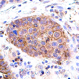 View Larger
EGFR in Human Lung Cancer. EGFR was detected in immersion fixed frozen sections of human lung cancer using Rabbit Anti-Human Phospho-EGFR (Y1173) Antigen Affinity-purified Polyclonal Antibody (Catalog # AF1095) at 0.5 µg/mL for 1 hour at room temperature followed by incubation with the Anti-Rabbit IgG VisUCyte™ HRP Polymer Antibody (VC003). Before incubation with the primary antibody, tissue was subjected to heat-induced epitope retrieval using Antigen Retrieval Reagent-Basic (CTS013). Tissue was stained using DAB (brown) and counterstained with hematoxylin (blue). Specific staining was localized to cell membrane in cancer cells. Staining was performed using our protocol for IHC Staining with VisUCyte HRP Polymer Detection Reagents.
 antibody in A431 Human Cell Line antibody by Flow Cytometry.") View Larger
View Larger
Detection of Phospho-EGFR (Y1173) in A431 Human Cell Line by Flow Cytometry. A431 human epithelial carcinoma cells were untreated (open histogram), or treated for 5 minutes with 100 ng/mL Recombinant Human EGF (Catalog # 236-EG; filled histogram) then stained with Rabbit Anti-Human Phospho-EGFR (Y1173) Antigen Affinity-purified Polyclonal Antibody (Catalog # AF1095), followed by Phycoerythrin-conjugated Anti-Rabbit IgG Secondary Antibody (Catalog # F0110). To facilitate intracellular staining, cells were fixed with para-formaldehyde and permeabilized with saponin.
 antibody by Simple Western<sup>TM</sup>.") View Larger
View Larger
Detection of Human Phospho-EGFR (Y1173) by Simple WesternTM. Simple Western lane view shows lysates of A431 human epithelial carcinoma cell line untreated (-) or treated (+) with 10 ng/mL Recombinant Human EGF (Catalog # 236-EG) for 5 minutes, loaded at 0.2 mg/mL. A specific band was detected for Phospho-EGFR (Y1173) at approximately 265 kDa (as indicated) using 2 µg/mL of Rabbit Anti-Human Phospho-EGFR (Y1173) Antigen Affinity-purified Polyclonal Antibody (Catalog # AF1095). This experiment was conducted under reducing conditions and using the 66-440 kDa separation system.
Reconstitution Calculator
Preparation and Storage
- 12 months from date of receipt, -20 to -70 °C as supplied.
- 1 month, 2 to 8 °C under sterile conditions after reconstitution.
- 6 months, -20 to -70 °C under sterile conditions after reconstitution.
Background: EGFR
Epidermal growth factor receptor (EGFR, also known as ErbB1 and HER1) is the founding member of the ErbB family of receptor tyrosine kinases. Ligand binding induces receptor dimerization and autophosphorylation on multiple tyrosine residues. Phosphorylation of Y1173 creates a binding site for the protein tyrosine phosphatase SHP-1, leading to attenuation of receptor signaling.
Product Datasheets
Citations for Human Phospho-EGFR (Y1173) Antibody
R&D Systems personnel manually curate a database that contains references using R&D Systems products. The data collected includes not only links to publications in PubMed, but also provides information about sample types, species, and experimental conditions.
7
Citations: Showing 1 - 7
Filter your results:
Filter by:
-
DNA damage alters EGFR signaling and reprograms cellular response via Mre-11
Authors: Y Volman, R Hefetz, E Galun, J Rachmilewi
Scientific Reports, 2022-04-06;12(1):5760.
Species: Human
Sample Types: Cell Lysates
Applications: Western Blot -
EGFR phosphorylates HDAC1 to regulate its expression and anti-apoptotic function
Authors: S Bahl, H Ling, NPN Acharige, I Santos-Bar, MKH Pflum, E Seto
Cell Death & Disease, 2021-05-11;12(5):469.
Species: Human
Sample Types: Cell Lysates, Whole Cells
Applications: Flow Cytometry, IP, Western Blot -
ADAM17 substrate release in proximal tubule drives kidney fibrosis
JCI Insight, 2016-08-18;1(13):.
Species: Mouse
Sample Types: Whole Tissue
Applications: IHC -
Phenotypic diversity of breast cancer-related mutations in metalloproteinase-disintegrin ADAM12.
Authors: Qi, Yue, Duhachek-Muggy, Sara, Li, Hui, Zolkiewska, Anna
PLoS ONE, 2014-03-20;9(3):e92536.
Species: Human
Sample Types: Cell Culture Supernates
Applications: Western Blot -
Analysis of Epithelial Growth Factor-Receptor (EGFR) Phosphorylation in Uterine Smooth Muscle Tumors: Correlation to Mucin-1 and Galectin-3 Expression.
Authors: Weissenbacher, Tobias, Vrekoussis, Thomas, Roeder, David, Makrigiannakis, Antonis, Mayr, Doris, Ditsch, Nina, Friese, Klaus, Jeschke, Udo, Dian, Darius
Int J Mol Sci, 2013-02-28;14(3):4783-92.
Species: Human
Sample Types: Whole Tissue
Applications: IHC-P -
Repulsion of cerebellar granule neurons by chondroitin sulfate proteoglycans is mediated by MAPK pathway.
Authors: Kaneko M, Kubo T, Hata K, Yamaguchi A, Yamashita T
Neurosci. Lett., 2007-07-05;423(1):62-7.
Species: Mouse
Sample Types: Cell Lysates
Applications: Western Blot -
Protein C is an autocrine growth factor for human skin keratinocytes.
Authors: Xue M, Campbell D, Jackson CJ
J. Biol. Chem., 2007-02-09;282(18):13610-6.
Species: Human
Sample Types: Whole Tissue
Applications: IHC-P
FAQs
No product specific FAQs exist for this product, however you may
View all Antibody FAQsReviews for Human Phospho-EGFR (Y1173) Antibody
There are currently no reviews for this product. Be the first to review Human Phospho-EGFR (Y1173) Antibody and earn rewards!
Have you used Human Phospho-EGFR (Y1173) Antibody?
Submit a review and receive an Amazon gift card.
$25/€18/£15/$25CAN/¥75 Yuan/¥2500 Yen for a review with an image
$10/€7/£6/$10 CAD/¥70 Yuan/¥1110 Yen for a review without an image


