Human Phospho-Histone H2AX (S139) Antibody Summary
Applications
Please Note: Optimal dilutions should be determined by each laboratory for each application. General Protocols are available in the Technical Information section on our website.
Scientific Data
 View Larger
Histone H2AX in Human Breast Cancer Tissue. Histone H2AX was detected in immersion fixed paraffin-embedded sections of human breast cancer tissue using Rabbit Anti-Human Phospho-Histone H2AX (S139) Antigen Affinity-purified Polyclonal Antibody (Catalog # AF2288) at 10 µg/mL overnight at 4 °C. Before incubation with the primary antibody tissue was subjected to heat-induced epitope retrieval using Antigen Retrieval Reagent-Basic (Catalog # CTS013). Tissue was stained using the Anti-Rabbit HRP-DAB Cell & Tissue Staining Kit (brown; Catalog # CTS005) and counterstained with hematoxylin (blue). View our protocol for Chromogenic IHC Staining of Paraffin-embedded Tissue Sections.
 View Larger
Detection of Human Phospho-Histone H2AX (S139) by Western Blot. Western blot shows lysates of Jurkat human acute T cell leukemia cell line untreated (-) or treated (+) with 1 µM camptothecin (CPT) for for the indicated times. PVDF membrane was probed with 0.5 µg/mL of Rabbit Anti-Human Phospho-Histone H2AX (S139) Antigen Affinity-purified Polyclonal Antibody (Catalog # AF2288), followed by HRP-conjugated Anti-Rabbit IgG Secondary Antibody (Catalog # HAF008). A specific band was detected for Phospho-Histone H2AX (S139) at approximately 20 kDa (as indicated). This experiment was conducted under reducing conditions and using Immunoblot Buffer Group 1.
 View Larger
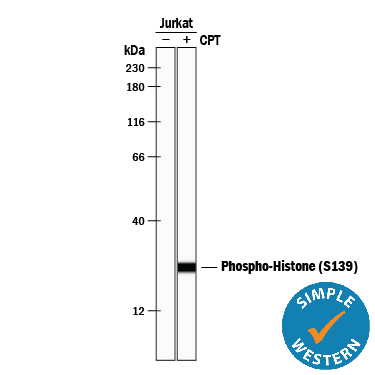
Detection of Human Phospho-Histone H2AX (S139) by Simple WesternTM. Simple Western lane view shows lysates of Jurkat human acute T cell leukemia cell line untreated (-) or treated (+) with 1 µM Camptothecin (CPT) for 24 hours, loaded at 0.2 mg/mL. A specific band was detected for Phospho-Histone H2AX (S139) at approximately 25 kDa (as indicated) using 5 µg/mL of Rabbit Anti-Human Phospho-Histone H2AX (S139) Antigen Affinity-purified Polyclonal Antibody (Catalog # AF2288). This experiment was conducted under reducing conditions and using the 12-230 kDa separation system.
 View Larger
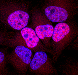
Phospho-Histone H2AX (S139) in HeLa Human Cell Line. Histone H2AX phosphorylated at S139 was detected in immersion fixed HeLa human cervical epithelial carcinoma cell line treated with ultraviolet radiation using Rabbit Anti-Human Phospho-Histone H2AX (S139) Antigen Affinity-purified Polyclonal Antibody (Catalog # AF2288) at 10 µg/mL for 3 hours at room temperature. Cells were stained using the NorthernLights™ 557-conjugated Anti-Rabbit IgG Secondary Antibody (red; Catalog # NL004) and counterstained with DAPI (blue). Specific staining was localized to nuclei. View our protocol for Fluorescent ICC Staining of Cells on Coverslips.
Reconstitution Calculator
Preparation and Storage
- 12 months from date of receipt, -20 to -70 °C as supplied.
- 1 month, 2 to 8 °C under sterile conditions after reconstitution.
- 6 months, -20 to -70 °C under sterile conditions after reconstitution.
Background: Histone H2AX
Histone H2AX is phosphorylated at S139 in cells exposed to DNA double-strand break-inducing agents, such as ionizing radiation. The S139 phosphorylated H2AX, termed ( gamma -H2AX, marks the site of DNA double-strand breaks and serves to recruit cell cycle checkpoint and DNA repair factors to the site of damage.
Product Datasheets
Citations for Human Phospho-Histone H2AX (S139) Antibody
R&D Systems personnel manually curate a database that contains references using R&D Systems products. The data collected includes not only links to publications in PubMed, but also provides information about sample types, species, and experimental conditions.
17
Citations: Showing 1 - 10
Filter your results:
Filter by:
-
Cardiac radiotherapy induces electrical conduction reprogramming in the absence of transmural fibrosis
Authors: DM Zhang, R Navara, T Yin, J Szymanski, U Goldsztejn, C Kenkel, A Lang, C Mpoy, CE Lipovsky, Y Qiao, S Hicks, G Li, KMS Moore, C Bergom, BE Rogers, CG Robinson, PS Cuculich, JK Schwarz, SL Rentschler
Nature Communications, 2021-09-24;12(1):5558.
Species: Mouse
Sample Types: Whole Tissue
Applications: IHC -
Selective killing of homologous recombination-deficient cancer cell lines by inhibitors of the RPA:RAD52 protein-protein interaction
Authors: M Al-Mugotir, JJ Lovelace, J George, M Bessho, D Pal, L Struble, C Kolar, S Rana, A Natarajan, T Bessho, GEO Borgstahl
PLoS ONE, 2021-03-30;16(3):e0248941.
Species: Human
Sample Types: Cell Lysates
Applications: Western Blot -
Melflufen, a peptide-conjugated alkylator, is an efficient anti-neo-plastic drug in breast cancer cell lines
Authors: A Schepsky, G Asta Traus, J Petur Joel, S Ingthorsso, J Kricker, J Thor Bergt, A Asbjarnars, T Gudjonsson, N Nupponen, A Slipicevic, F Lehmann, T Gudjonsson
Cancer Med, 2020-07-27;0(0):.
Species: Human
Sample Types: Whole Cells
Applications: ICC -
Radiation Response of Murine Embryonic Stem Cells
Authors: CE Hellweg, V Shinde, SP Srinivasan, M Henry, T Rotshteyn, C Baumstark-, C Schmitz, S Feles, LF Spitta, R Hemmersbac, J Hescheler, A Sachinidis
Cells, 2020-07-09;9(7):.
Species: Mouse
Sample Types: Whole Cells
Applications: ICC -
Evaluation of the role of mitochondria in the non-targeted effects of ionizing radiation using cybrid cellular models
Authors: S Miranda, M Correia, AG Dias, A Pestana, P Soares, J Nunes, J Lima, V Máximo, P Boaventura
Sci Rep, 2020-04-09;10(1):6131.
Species: Human
Sample Types: Whole Cells
Applications: ICC -
BRCA1 intronic Alu elements drive gene rearrangements and PARP inhibitor resistance
Authors: Y Wang, AJ Bernhardy, J Nacson, JJ Krais, YF Tan, E Nicolas, MR Radke, E Handorf, A Llop-Gueva, J Balmaña, EM Swisher, V Serra, S Peri, N Johnson
Nat Commun, 2019-12-11;10(1):5661.
Species: Human
Sample Types: Whole Cells
Applications: ICC -
PTEN expression in U251 glioma cells enhances their sensitivity to ionizing radiation by suppressing DNA repair capacity
Authors: HL Li, CY Wang, J Fu, XJ Yang, Y Sun, YH Shao, LH Zhang, XM Yang, XL Zhang, J Lin
Eur Rev Med Pharmacol Sci, 2019-12-01;23(23):10453-10458.
Species: Human
Sample Types: Whole Cells
Applications: ICC -
Arginine methyltransferase PRMT8 provides cellular stress tolerance in aging motoneurons
Authors: Z Simandi, K Pajer, K Karolyi, T Sieler, LL Jiang, Z Kolostyak, Z Sari, Z Fekecs, A Pap, A Patsalos, GA Contreras, B Reho, Z Papp, X Guo, A Horvath, G Kiss, Z Keresztess, G Vámosi, J Hickman, H Xu, D Dormann, T Hortobagyi, M Antal, A Nogradi, L Nagy
J. Neurosci., 2018-07-27;0(0):.
Species: Mouse
Sample Types: Cell Lysates, Whole Tissue
Applications: IHC, Western Blot -
SMN deficiency in severe models of spinal muscular atrophy causes widespread intron retention and DNA damage
Authors: M Jangi, C Fleet, P Cullen, SV Gupta, S Mekhoubad, E Chiao, N Allaire, CF Bennett, F Rigo, AR Krainer, JA Hurt, JP Carulli, JF Staropoli
Proc. Natl. Acad. Sci. U.S.A, 2017-03-07;0(0):.
Species: Mouse
Sample Types: Whole Tissue
Applications: IHC -
Synergistic Activity of Deguelin and Fludarabine in Cells from Chronic Lymphocytic Leukemia Patients and in the New Zealand Black Murine Model
Authors: N Rebolleda, I Losada-Fer, G Perez-Chac, R Castejon, S Rosado, M Morado, MT Vallejo-Cr, A Martinez, JA Vargas-Nuñ, P Perez-Acie
PLoS ONE, 2016-04-21;11(4):e0154159.
Species: Human
Sample Types: Cell Lysates
Applications: Western Blot -
Insulin and IGF1 signalling pathways in human astrocytes in vitro and in vivo; characterisation, subcellular localisation and modulation of the receptors.
Authors: Garwood C, Ratcliffe L, Morgan S, Simpson J, Owens H, Vazquez-Villasenor I, Heath P, Romero I, Ince P, Wharton S
Mol Brain, 2015-08-22;8(0):51.
-
Cyclin-dependent kinase five mediates activation of lung xanthine oxidoreductase in response to hypoxia.
Authors: Kim, Bo S, Serebreni, Leonid, Fallica, Jonathan, Hamdan, Omar, Wang, Lan, Johnston, Laura, Kolb, Todd, Damarla, Mahendra, Damico, Rachel, Hassoun, Paul M
PLoS ONE, 2015-04-01;10(4):e0124189.
Species: Rat
Sample Types: Cell Lysates
Applications: Western Blot -
Inhibition of CHK1 kinase by Go6976 converts 8-chloro-adenosine-induced G2/M arrest into S arrest in human myelocytic leukemia K562 cells.
Authors: Jia XZ, Yang SY, Zhou J, Li SY, Ni JH, An GS, Jia HT
Biochem. Pharmacol., 2008-11-18;77(5):770-80.
Species: Human
Sample Types: Cell Lysates, Whole Cells
Applications: ICC, Western Blot -
Inhibition of topoisomerase II by 8-chloro-adenosine triphosphate induces DNA double-stranded breaks in 8-chloro-adenosine-exposed human myelocytic leukemia K562 cells.
Authors: Yang SY, Jia XZ, Feng LY, Li SY, An GS, Ni JH, Jia HT
Biochem. Pharmacol., 2008-10-28;77(3):433-43.
Species: Human
Sample Types: Cell Lysates, Whole Cells
Applications: ICC, Western Blot -
DNA cross-linking, double-strand breaks, and apoptosis in corneal endothelial cells after a single exposure to mitomycin C.
Authors: Roh DS, Cook AL, Rhee SS, Joshi A, Kowalski R, Dhaliwal DK, Funderburgh JL
Invest. Ophthalmol. Vis. Sci., 2008-07-24;49(11):4837-43.
Species: Goat
Sample Types: Whole Cells
Applications: ICC -
Induction of MHC class I-related chain B (MICB) by 5-aza-2'-deoxycytidine.
Authors: Tang KF, He CX, Zeng GL, Wu J, Song GB, Shi YS, Zhang WG, Huang AL, Steinle A, Ren H
Biochem. Biophys. Res. Commun., 2008-04-04;370(4):578-83.
Species: Human
Sample Types: Whole Cells
Applications: ICC -
Histone H2AX and Fanconi anemia FANCD2 function in the same pathway to maintain chromosome stability.
Authors: Bogliolo M, Lyakhovich A, Callen E, Castella M, Cappelli E, Ramirez MJ, Creus A, Marcos R, Kalb R, Neveling K, Schindler D, Surralles J
EMBO J., 2007-02-15;26(5):1340-51.
Species: Human
Sample Types: Whole Cells
Applications: ICC
FAQs
No product specific FAQs exist for this product, however you may
View all Antibody FAQsReviews for Human Phospho-Histone H2AX (S139) Antibody
There are currently no reviews for this product. Be the first to review Human Phospho-Histone H2AX (S139) Antibody and earn rewards!
Have you used Human Phospho-Histone H2AX (S139) Antibody?
Submit a review and receive an Amazon gift card.
$25/€18/£15/$25CAN/¥75 Yuan/¥2500 Yen for a review with an image
$10/€7/£6/$10 CAD/¥70 Yuan/¥1110 Yen for a review without an image

