Human Phospho-Paxillin (Y31) Antibody Summary
Applications
Please Note: Optimal dilutions should be determined by each laboratory for each application. General Protocols are available in the Technical Information section on our website.
Scientific Data
 View Larger
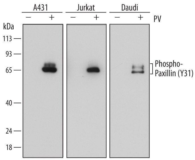
Detection of Human Phospho-Paxillin (Y31) by Western Blot. Western blot shows lysates of A431 human epithelial carcinoma cell line, Jurkat human acute T cell leukemia cell line, and Daudi human Burkitt's lymphoma cell line untreated (-) or treated (+) with 1 mM Pervanadate (PV) for 30 minutes. PVDF membrane was probed with 0.1 µg/mL of Mouse Anti-Human Phospho-Paxillin (Y31) Antigen Affinity-purified Monoclonal Antibody (Catalog # MAB61641) followed by HRP-conjugated Anti-Mouse IgG Secondary Antibody (Catalog # HAF007). Specific bands were detected for Phospho-Paxillin (Y31) at approximately 65 to 68 kDa (as indicated). This experiment was conducted under reducing conditions and using Immunoblot Buffer Group 1.
 View Larger
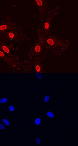
Paxillin in HUVEC Human Cells. Paxillin phosphorylated at Y31 was detected in immersion fixed HUVEC human umbilical vein endothelial cells using Mouse Anti-Human Phospho-Paxillin (Y31) Antigen Affinity-purified Monoclonal Antibody (Catalog # MAB61641) at 10 µg/mL for 3 hours at room temperature. Cells were stained using the NorthernLights™ 557-conjugated Anti-Mouse IgG Secondary Antibody (red, upper panel; Catalog # NL007) and counterstained with DAPI (blue, lower panel). Specific staining was localized to nuclei and focal adhesions. View our protocol for Fluorescent ICC Staining of Cells on Coverslips.
Reconstitution Calculator
Preparation and Storage
- 12 months from date of receipt, -20 to -70 °C as supplied.
- 1 month, 2 to 8 °C under sterile conditions after reconstitution.
- 6 months, -20 to -70 °C under sterile conditions after reconstitution.
Background: Paxillin
Paxillin is a 65 kDa cytoskeletal adaptor protein and member of the Paxillin family. Human Paxillin is 591 amino acids (aa) in length and contains four LIM zinc-binding domains. Alternative splicing produces three isoforms. Human Paxillin shares 94% and 85% aa identity with mouse and rat Paxillin, respectively. Paxillin is found at the interface between actin filaments and the plasma membrane, and it localizes to focal adhesions, where it provides a platform for the integration and coordination of adhesion- and growth factor-related signals. Paxillin phosphorylation at tyrosines 31 and 118 is required for integrin-mediated cytoskeletal reorganization, and may play a role in the disassembly of focal adhesions and stress fibers during cellular transformation.
Product Datasheets
FAQs
No product specific FAQs exist for this product, however you may
View all Antibody FAQsReviews for Human Phospho-Paxillin (Y31) Antibody
There are currently no reviews for this product. Be the first to review Human Phospho-Paxillin (Y31) Antibody and earn rewards!
Have you used Human Phospho-Paxillin (Y31) Antibody?
Submit a review and receive an Amazon gift card.
$25/€18/£15/$25CAN/¥75 Yuan/¥2500 Yen for a review with an image
$10/€7/£6/$10 CAD/¥70 Yuan/¥1110 Yen for a review without an image


