Human Phospho-STAT6 (Y641) Antibody Summary
Accession # P42226
Applications
Please Note: Optimal dilutions should be determined by each laboratory for each application. General Protocols are available in the Technical Information section on our website.
Scientific Data
 View Larger
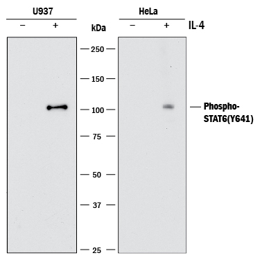
Detection of Human Phospho-STAT6 by Western Blot. Western blot shows lysates of U937 human histiocytic lymphoma cell line and HeLa human cervical epithelial carcinoma cell line untreated (-) or treated (+) with 100 ng/mL Recombinant Human IL-4 (Catalog # 204-IL) for 15 minutes or 2 ng/mL Recombinant Human IL-4 (Catalog # 204-IL) for 20 minutes, respectively. PVDF membrane was probed with 1 µg/mL of Rabbit Anti-Human Phospho-STAT6 (Y641) Monoclonal Antibody (Catalog # MAB3717) followed by HRP-conjugated Anti-Rabbit IgG Secondary Antibody (Catalog # HAF008). A specific band was detected for STAT6 at approximately 100 kDa (as indicated). This experiment was conducted under reducing conditions and using Immunoblot Buffer Group 1.
 View Larger
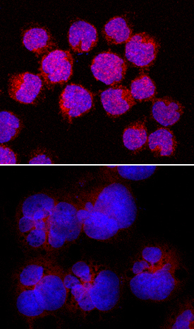
Phospho-STAT6 (Y641) in Daudi Human Cell Line. STAT6 phosphorylated at Y641 was detected in immersion fixed Daudi human Burkitt's lymphoma cell line, untreated (lower panel) or treated with Recombinant Human IL-4 (Catalog # 204-IL; upper panel) using Rabbit Anti-Human Phospho-STAT6 (Y641) Monoclonal Antibody (Catalog # MAB3717) at 5 µg/mL for 3 hours at room temperature. Cells were stained using the NorthernLights™ 557-conjugated Anti-Rabbit IgG Secondary Antibody (red; Catalog # NL004) and counterstained with DAPI (blue). Specific staining was localized to cytoplasm. View our protocol for Fluorescent ICC Staining of Non-adherent Cells.
 View Larger
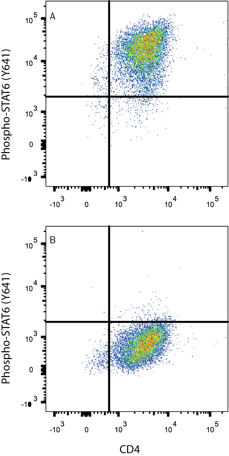
Detection of STAT6 in Human peripheral blood mononuclear cells (PBMCs) by Flow Cytometry. Human peripheral blood mononuclear cells (PBMCs) either (A) Th2-stimulated or (B) unstimulated were stained with Rabbit Anti-Human Phospho-STAT6 (Y641) Monoclonal Antibody (Catalog # MAB3717) followed by anti-Rabbit IgG PE-conjugated secondary antibody (Catalog # F0110) and Mouse Anti-Human CD4 APC-conjugated Monoclonal Antibody (Catalog # FAB3791A). Quadrant markers were set based on control antibody staining (Catalog # MAB1050). To facilitate intracellular staining, cells were fixed with Flow Cytometry Fixation Buffer (Catalog # FC004) and permeabilized with methanol. View our protocol for Staining Intracellular Molecules.
Reconstitution Calculator
Preparation and Storage
- 12 months from date of receipt, -20 to -70 °C as supplied.
- 1 month, 2 to 8 °C under sterile conditions after reconstitution.
- 6 months, -20 to -70 °C under sterile conditions after reconstitution.
Background: STAT6
Signal Transducer and Activator of Transcription 6 (STAT6) mediates the signaling of cytokines such as IL-4 and IL-13. STAT6 acts as a signal transducer in the cytoplasm and, upon phosphorylation at Y641, translocates to the nucleus and binds to the DNA consensus site TTCN4GAA. Knockout studies in mice suggest that STAT6 functions in differentiation of T helper 2 (Th2) cells, expression of cell surface markers, and class switch of immunoglobulins.
Product Datasheets
FAQs
No product specific FAQs exist for this product, however you may
View all Antibody FAQsReviews for Human Phospho-STAT6 (Y641) Antibody
There are currently no reviews for this product. Be the first to review Human Phospho-STAT6 (Y641) Antibody and earn rewards!
Have you used Human Phospho-STAT6 (Y641) Antibody?
Submit a review and receive an Amazon gift card.
$25/€18/£15/$25CAN/¥75 Yuan/¥2500 Yen for a review with an image
$10/€7/£6/$10 CAD/¥70 Yuan/¥1110 Yen for a review without an image


