Human/Porcine/Canine CD90/Thy1 Antibody Summary
Applications
Please Note: Optimal dilutions should be determined by each laboratory for each application. General Protocols are available in the Technical Information section on our website.
Scientific Data
 View Larger
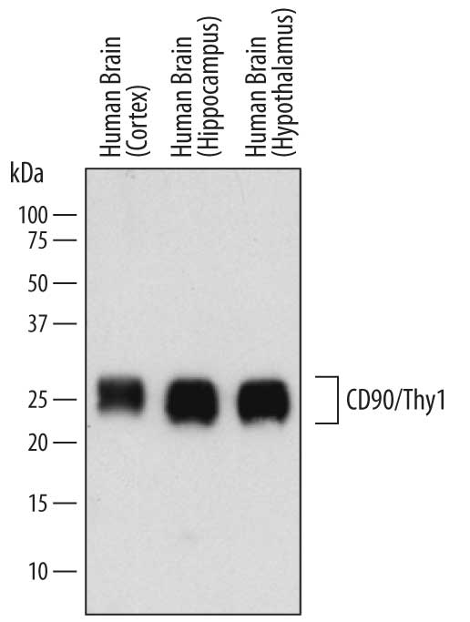
Detection of Human CD90/Thy1 by Western Blot. Western blot shows lysates of human brain (cortex) tissue, human brain (hippocampus) tissue, and human brain (hypothalamus) tissue. PVDF membrane was probed with 1 µg/mL of Sheep Anti-Human/Porcine/Canine CD90/Thy1 Antigen Affinity-purified Polyclonal Antibody (Catalog # AF2067) followed by HRP-conjugated Anti-Sheep IgG Secondary Antibody (Catalog # HAF016). A specific band was detected for CD90/Thy1 at approximately 22-30 kDa (as indicated). This experiment was conducted under reducing conditions and using Immunoblot Buffer Group 1.
 View Larger
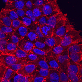
CD90/Thy1 in BG01V Human Embyonic Stem Cells. CD90/Thy1 was detected in immersion fixed BG01V human embryonic stem cells using Sheep Anti-Human/Porcine/Canine CD90/Thy1 Antigen Affinity-purified Polyclonal Antibody (Catalog # AF2067) at 10 µg/mL for 3 hours at room temperature. Cells were stained using the NorthernLights™ 557-conjugated Anti-Sheep IgG Secondary Antibody (red; Catalog # NL010) and counterstained with DAPI (blue). Specific staining was localized to cytoplasm. View our protocol for Fluorescent ICC Staining of Stem Cells on Coverslips.
 View Larger
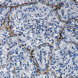
CD90/Thy1 in Human Prostate Cancer Tissue. CD90/Thy1 was detected in formalin fixed paraffin-embedded sections of human prostate cancer tissue using Sheep Anti-Human/Porcine/Canine CD90/Thy1 Antigen Affinity-purified Polyclonal Antibody (Catalog # AF2067) at 1.7 µg/mL overnight at 4 °C. Tissue was stained using the Anti-Sheep HRP-DAB Cell & Tissue Staining Kit (brown; Catalog # CTS019) and counterstained with hematoxylin (blue). Specific staining was localized to endothelial cells. View our protocol for Chromogenic IHC Staining of Paraffin-embedded Tissue Sections.
 View Larger
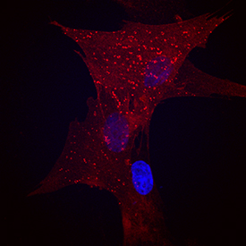
CD90/Thy1 in Canine Mesenchymal Stem Cells. CD90/Thy1 was detected in immersion fixed canine mesenchymal stem cells using Sheep Anti-Human/Porcine/Canine CD90/Thy1 Antigen Affinity-purified Polyclonal Antibody (Catalog # AF2067) at 10 µg/mL for 3 hours at room temperature. Cells were stained using the NorthernLights™ 557-conjugated Anti-Sheep IgG Secondary Antibody (red; Catalog # NL010) and counterstained with DAPI (blue). Specific staining was localized to cell surfaces. View our protocol for Fluorescent ICC Staining of Stem Cells on Coverslips.
 View Larger
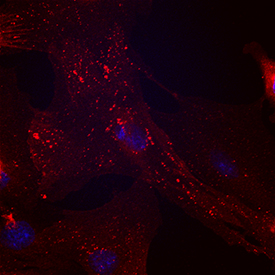
CD90/Thy1 in Porcine Mesenchymal Stem Cells. CD90/Thy1 was detected in immersion fixed porcine mesenchymal stem cells using Sheep Anti-Human/Porcine/Canine CD90/Thy1 Antigen Affinity-purified Polyclonal Antibody (Catalog # AF2067) at 10 µg/mL for 3 hours at room temperature. Cells were stained using the NorthernLights™ 557-conjugated Anti-Sheep IgG Secondary Antibody (red; Catalog # NL010) and counterstained with DAPI (blue). Specific staining was localized to cell surfaces and cytoplasm. View our protocol for Fluorescent ICC Staining of Stem Cells on Coverslips.
 View Larger
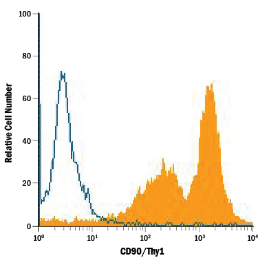
Detection of CD90/Thy1 in Human Mesenchymal Stem Cells by Flow Cytometry. Human mesenchymal stem cells were stained with Sheep Anti-Human/Porcine/Canine CD90/Thy1 Antigen Affinity-purified Polyclonal Antibody (Catalog # AF2067, filled histogram) or isotype control antibody (Catalog # 5-001-A, open histogram), followed by PE-conjugated Anti-Sheep IgG Secondary Antibody (Catalog # F0126).
 View Larger
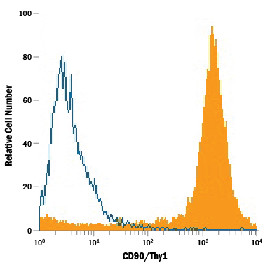
Detection of CD90/Thy1 in Porcine Mesenchymal Stem Cells by Flow Cytometry. Porcine mesenchymal stem cells were stained with Sheep Anti-Human/Porcine/Canine CD90/Thy1 Antigen Affinity-purified Polyclonal Antibody (Catalog # AF2067, filled histogram) or isotype control antibody (Catalog # 5-001-A, open histogram), followed by PE-conjugated Anti-Sheep IgG Secondary Antibody (Catalog # F0126).
 View Larger
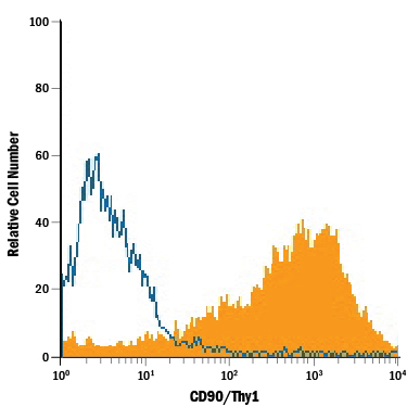
Detection of CD90/Thy1 in Canine Mesenchymal Stem Cells by Flow Cytometry. Canine mesenchymal stem cells were stained with Sheep Anti-Human/Porcine/Canine CD90/Thy1 Antigen Affinity-purified Polyclonal Antibody (Catalog # AF2067, filled histogram) or isotype control antibody (Catalog # 5-001-A, open histogram), followed by PE-conjugated Anti-Sheep IgG Secondary Antibody (Catalog # F0126).
Reconstitution Calculator
Preparation and Storage
- 12 months from date of receipt, -20 to -70 degreesC as supplied. 1 month, 2 to 8 degreesC under sterile conditions after reconstitution. 6 months, -20 to -70 degreesC under sterile conditions after reconstitution.
Background: CD90/Thy1
CD90 (also Thy1/Thymus cell antigen 1) is a 25-35 kDa glycoprotein member of the Immunoglobulin superfamily of molecules. It is expressed on neurons, ovarian follicular cells, endothelial cells, fibroblasts and circulating CD34 + stem cells, and appears to act as an adhesion molecule. CD90 is known to bind to integrins beta 2, beta 5 and beta 3, the latter often accompanied by additional binding to syndecan-4. In the postnatal nervous system, this inhibits neurite outgrowth while stabilizing newly formed axonal networks. In the vascular system, CD90 mediates the extravasation of leukocytes. And in lung, a CD90: alpha v beta 5 interaction inhibits the extracellular activation of latent TGF-beta. Mature human CD90 is a 111 amino acid (aa) GPI-linked protein (aa 20-130). It contains one V-type Ig-like domain (aa 20-126) with a heparin-binding motif (aa 56-60), and a GPI-anchor amidated cysteine at position 130. The integrin binding site consists of an Arg35LeuAsp37 tripeptide. CD90 apparently forms 50-60 kDa homodimers and 150 kDa homomultimers. There is one potential isoform variant that contains a 12 aa substitution for aa 1-13. Over aa 20-141, human CD90 shares 64% aa identity with mouse CD90 and 84% or 83% with horse and dog respectively.
Product Datasheets
Citations for Human/Porcine/Canine CD90/Thy1 Antibody
R&D Systems personnel manually curate a database that contains references using R&D Systems products. The data collected includes not only links to publications in PubMed, but also provides information about sample types, species, and experimental conditions.
6
Citations: Showing 1 - 6
Filter your results:
Filter by:
-
Improved Wound Healing and Skin Regeneration Ability of 3,2'-Dihydroxyflavone-Treated Mesenchymal Stem Cell-Derived Extracellular Vesicles
Authors: S Kim, Y Shin, Y Choi, KM Lim, Y Jeong, AA Dayem, Y Lee, J An, K Song, SB Jang, SG Cho
International Journal of Molecular Sciences, 2023-04-09;24(8):.
Species: Human
Sample Types: Whole Cells
Applications: Flow Cytometry -
In vivo recellularization of xenogeneic vascular grafts decellularized with high hydrostatic pressure method in a porcine carotid arterial interpose model
Authors: S Kurokawa, Y Hashimoto, S Funamoto, K Murata, A Yamashita, K Yamazaki, T Ikeda, K Minatoya, A Kishida, H Masumoto
PLoS ONE, 2021-07-22;16(7):e0254160.
Species: Porcine
Sample Types: Whole Tissue
Applications: IHC -
Notch signalling drives synovial fibroblast identity and arthritis pathology
Authors: K Wei, I Korsunsky, JL Marshall, A Gao, GFM Watts, T Major, AP Croft, J Watts, PE Blazar, JK Lange, TS Thornhill, A Filer, K Raza, LT Donlin, CW Siebel, CD Buckley, S Raychaudhu, MB Brenner
Nature, 2020-04-22;582(7811):259-264.
Species: Human
Sample Types: Whole Tissue
Applications: IHC -
Novel chitosan/agarose/hydroxyapatite nanocomposite scaffold for bone tissue engineering applications: comprehensive evaluation of biocompatibility and osteoinductivity with the use of osteoblasts and mesenchymal stem cells
Authors: P Kazimiercz, A Benko, M Nocun, A Przekora
Int J Nanomedicine, 2019-08-19;14(0):6615-6630.
Species: Canine
Sample Types: Whole Cells
Applications: ICC -
Podoplanin regulates the migration of mesenchymal stromal cells and their interaction with platelets
Authors: LSC Ward, L Sheriff, JL Marshall, JE Manning, A Brill, GB Nash, HM McGettrick
J. Cell. Sci., 2019-02-25;0(0):.
Species: Human
Sample Types: Whole Tissue
Applications: IHC-Fr -
Pathological Remodeling of Mitral Valve Leaflets from Unphysiologic Leaflet Mechanics after Undersized Mitral Annuloplasty to Repair Ischemic Mitral Regurgitation
Authors: A Sielicka, EL Sarin, W Shi, F Sulejmani, D Corporan, K Kalra, VH Thourani, W Sun, RA Guyton, M Padala
J Am Heart Assoc, 2018-11-06;7(21):e009777.
Species: Porcine
Sample Types: Whole Tissue
Applications: IHC
FAQs
No product specific FAQs exist for this product, however you may
View all Antibody FAQsReviews for Human/Porcine/Canine CD90/Thy1 Antibody
There are currently no reviews for this product. Be the first to review Human/Porcine/Canine CD90/Thy1 Antibody and earn rewards!
Have you used Human/Porcine/Canine CD90/Thy1 Antibody?
Submit a review and receive an Amazon gift card.
$25/€18/£15/$25CAN/¥75 Yuan/¥2500 Yen for a review with an image
$10/€7/£6/$10 CAD/¥70 Yuan/¥1110 Yen for a review without an image


