Human PTEN Antibody Summary
Applications
Please Note: Optimal dilutions should be determined by each laboratory for each application. General Protocols are available in the Technical Information section on our website.
Scientific Data
 View Larger
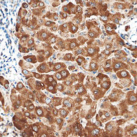
PTEN in Human Liver. PTEN was detected in immersion fixed paraffin-embedded sections of human liver using Goat Anti- Human PTEN Antigen Affinity-purified Polyclonal Antibody (Catalog # AF6655) at 10 µg/mL overnight at 4 °C. Before incubation with the primary antibody, tissue was subjected to heat-induced epitope retrieval using Antigen Retrieval Reagent-Basic (Catalog # CTS013). Tissue was stained using the Anti-Goat HRP-DAB Cell & Tissue Staining Kit (brown; Catalog # CTS008) and counterstained with hematoxylin (blue). Specific staining was localized to cytoplasm of hepatocytes. View our protocol for Chromogenic IHC Staining of Paraffin-embedded Tissue Sections.
 View Larger
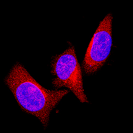
PTEN in Neuro‑2A Mouse Cell Line. PTEN was detected in immersion fixed Neuro-2A mouse neuroblastoma cell line using Goat Anti-Human PTEN Antigen Affinity-purified Polyclonal Antibody (Catalog # AF6655) at 1.7 µg/mL for 3 hours at room temperature. Cells were stained using the NorthernLights™ 557-conjugated Anti-Goat IgG Secondary Antibody (red; Catalog # NL001) and counterstained with DAPI (blue). Specific staining was localized to cytoplasm. View our protocol for Fluorescent ICC Staining of Cells on Coverslips.
Reconstitution Calculator
Preparation and Storage
- 12 months from date of receipt, -20 to -70 degreesC as supplied. 1 month, 2 to 8 degreesC under sterile conditions after reconstitution. 6 months, -20 to -70 degreesC under sterile conditions after reconstitution.
Background: PTEN
The tumor suppressor gene PTEN (phosphatase and tensin homolog deleted on chromosome 10), also known as MMAC1 (mutated in multiple advanced cancers 1), encodes a phosphatase that contains the catalytic signature motif (HCXXGXXRS/T) found in all members of the protein tyrosine phosphatase family. In vitro, the recombinant PTEN has both lipid phosphatase and protein phosphatase activities (1, 2). Interestingly, accumulating evidence has shown that the tumor suppressor activity of PTEN relies on its ability to dephosphorylate phosphatidylinositol (3, 4, 5)-triphosphate specifically at position 3 of the inositol ring (3). This activity reduces the levels of phosphatidylinositol (3, 4, 5)-triphosphate which is specifically produced from phosphatidylinositol (4, 5)-diphosphate by PI 3-kinase upon activation by a variety of stimuli. Therefore, PTEN antagonizes PI 3-kinase-induced downstream signaling events and cellular processes including cell growth, apoptosis and cell motility. In vivo, the importance of PTEN catalytic activity in its tumor suppressor functions is underscored by the fact that the majority of PTEN missense mutations detected in tumor specimens target the phosphatase domain and cause a loss in PTEN phosphatase activity (4).
- Maehama, T. and J. Dixon (1998) J. Biol. Chem. 273:13375.
- Das, S. et al. (2003) Proc. Natl. Acad. Sci. USA 100:7491.
- Myers, M. et al. (1998) Proc. Natl. Acad. Sci. USA 95:13513.
- Waite, K. and C. Eng (2002) Am. J. Hum. Genet. 70:829.
Product Datasheets
Product Specific Notices
This product is covered by the following U.S. patent: USSN # 10/299,003.FAQs
No product specific FAQs exist for this product, however you may
View all Antibody FAQsReviews for Human PTEN Antibody
Average Rating: 5 (Based on 1 Review)
Have you used Human PTEN Antibody?
Submit a review and receive an Amazon gift card.
$25/€18/£15/$25CAN/¥75 Yuan/¥2500 Yen for a review with an image
$10/€7/£6/$10 CAD/¥70 Yuan/¥1110 Yen for a review without an image
Filter by:


