Human Purine Nucleoside Phosphorylase/PNP Antibody
Human Purine Nucleoside Phosphorylase/PNP Antibody Summary
Met1-Ser289
Accession # P00491
Applications
Please Note: Optimal dilutions should be determined by each laboratory for each application. General Protocols are available in the Technical Information section on our website.
Scientific Data
 View Larger
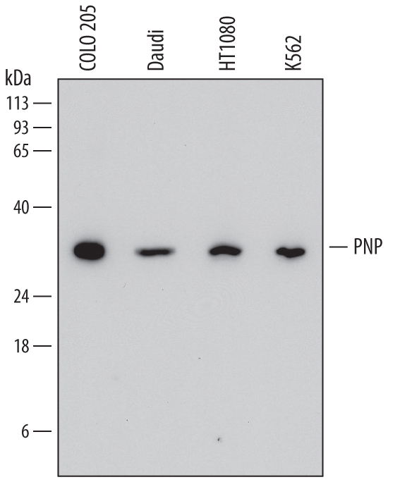
Detection of Human Purine Nucleoside Phosphorylase/PNP by Western Blot. Western blot shows lysates of COLO 205 human colorectal adenocarcinoma cell line, Daudi human Burkitt's lymphoma cell line, HT1080 human fibrosarcoma cell line, and K562 human chronic myelogenous leukemia cell line. PVDF membrane was probed with 0.5 µg/mL of Mouse Anti-Human Purine Nucleoside Phosphorylase/PNP Monoclonal Antibody (Catalog # MAB6486) followed by HRP-conjugated Anti-Mouse IgG Secondary Antibody (Catalog # HAF018). A specific band was detected for Purine Nucleoside Phosphorylase/PNP at approximately 32 kDa (as indicated). This experiment was conducted under reducing conditions and using Immunoblot Buffer Group 1.
 View Larger
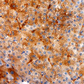
Purine Nucleoside Phosphorylase/PNP in Human Liver. Purine Nucleoside Phosphorylase/PNP was detected in immersion fixed paraffin-embedded sections of human liver using Mouse Anti-Human Purine Nucleoside Phosphorylase/PNP Monoclonal Antibody (Catalog # MAB6486) at 25 µg/mL overnight at 4 °C. Tissue was stained using the Anti-Mouse HRP-DAB Cell & Tissue Staining Kit (brown; Catalog # CTS002) and counter-stained with hematoxylin (blue). Specific staining was localized to endothelial cells in bile canaliculi. View our protocol for Chromogenic IHC Staining of Paraffin-embedded Tissue Sections.
Reconstitution Calculator
Preparation and Storage
- 12 months from date of receipt, -20 to -70 °C as supplied.
- 1 month, 2 to 8 °C under sterile conditions after reconstitution.
- 6 months, -20 to -70 °C under sterile conditions after reconstitution.
Background: Purine Nucleoside Phosphorylase/PNP
Purine Nucleoside Phosphorylase (PNP) catalyzes the phophorolysis of N-ribosidic bonds of purine nucleosides and deoxynucleosides. Physiological substrates of PNP include inosine, guanosine, and 2'-deoxyguanosine, but not adenosine (1). PNP is expressed in most tissues, with markedly greater expression in lymphoid tissues. Genetic deficiencies of PNP result in severely compromised T‑lymphocyte function and neurologic dysfunction (2, 3). PNP is used in assays for the measurement of inorganic phosphate (4).
- Schramm, V.L. (1998) Annu. Rev. Biochem. 67:693.
- Stoop, W. et al. (1977) N. Eng. J. Med. 296:651.
- Markert, M.L. (1991) Immunodefic. Rev. 3:45.
- Webb, M.R. (1992) Proc. Natl. Acad. Sci. USA. 89:4884.
Product Datasheets
FAQs
No product specific FAQs exist for this product, however you may
View all Antibody FAQsReviews for Human Purine Nucleoside Phosphorylase/PNP Antibody
There are currently no reviews for this product. Be the first to review Human Purine Nucleoside Phosphorylase/PNP Antibody and earn rewards!
Have you used Human Purine Nucleoside Phosphorylase/PNP Antibody?
Submit a review and receive an Amazon gift card.
$25/€18/£15/$25CAN/¥75 Yuan/¥2500 Yen for a review with an image
$10/€7/£6/$10 CAD/¥70 Yuan/¥1110 Yen for a review without an image

