Human/Rat Brevican Antibody Summary
Asp23-Pro911
Accession # AAH27971
Applications
Please Note: Optimal dilutions should be determined by each laboratory for each application. General Protocols are available in the Technical Information section on our website.
Scientific Data
 View Larger
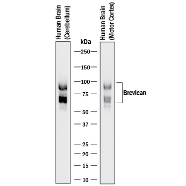
Detection of Human Brevican by Western Blot. Western blot shows lysates of human brain (cerebellum) tissue and human brain (motor cortex) tissue. PVDF membrane was probed with 0.1 µg/mL of Sheep Anti-Human/Rat Brevican Antigen Affinity-purified Polyclonal Antibody (Catalog # AF4009) followed by Donkey Anti-Sheep IgG HRP-conjugated Antigen Affinity-purified Polyclonal Antibody(Catalog # HAF016). Specific bands were detected for Brevican at approximately 60 and 90 kDa (as indicated). This experiment was conducted under reducing conditions and using Immunoblot Buffer Group 1.
 View Larger
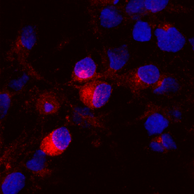
Brevican in Rat Cortical Stem Cells. Brevican was detected in immersion fixed rat cortical stem cells differentiated for 7 days by growth factor withdrawal using Sheep Anti-Human/Rat Brevican Antigen Affinity-purified Polyclonal Antibody (Catalog # AF4009) at 10 µg/mL for 3 hours at room temperature. Cells were stained using the NorthernLights™ 557-conjugated Anti-Sheep IgG Secondary Antibody (red; Catalog # NL010) and counterstained with DAPI (blue). Specific staining was localized to cytoplasm. View our protocol for Fluorescent ICC Staining of Stem Cells on Coverslips.
 View Larger
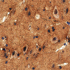
Brevican in Human Brain. Brevican was detected in immersion fixed paraffin-embedded sections of human brain (cortex) using Sheep Anti-Human/Rat Brevican Antigen Affinity-purified Polyclonal Antibody (Catalog # AF4009) at 5 µg/mL overnight at 4 °C. Tissue was stained using the Anti-Sheep HRP-DAB Cell & Tissue Staining Kit (brown; Catalog # CTS019) and counterstained with hematoxylin (blue). Specific staining was localized to neuropil. View our protocol for Chromogenic IHC Staining of Paraffin-embedded Tissue Sections.
Reconstitution Calculator
Preparation and Storage
- 12 months from date of receipt, -20 to -70 °C as supplied.
- 1 month, 2 to 8 °C under sterile conditions after reconstitution.
- 6 months, -20 to -70 °C under sterile conditions after reconstitution.
Background: Brevican
Brevican, also called BEHAB, is a secreted member of the the lectican family of proteoglycans that share a common domain structure (1). Brevican contains an Ig‑like V-set domain, two link domains, a Glu-rich region, a central region with glycosaminoglycan (GAG) modifications, an EGF-like domain, a C-type lectin domain, and a C-terminal Sushi/CRP-like domain (2). Brevican is restricted to the CNS and is expressed by astrocytes, oligodendrocytes, and neurons (3‑7). A GPI-anchored alternate splice form exists that is truncated following the central (GAG) region (2, 8). Brevican is cleaved by multiple proteases and exists in a number of distinct fragments (5, 9, 10). Full-length brevican consists of a 97 kDa core protein with up to approximately 100 kDa of attached chondroitin sulfate but not heparan sulfate chains (4, 7, 11, 12). Brevican associates with the extracellular matrix, perineuronal nets, and astrocyte cell surfaces by means of its chondroitin sulfate, GPI anchor, hyaluronic acid-binding link domains, and the core protein (4, 7, 8, 13). The secreted isoform is dominant during brain development and is up‑regulated in astrocytes following brain injury (2, 14). In human and rat, an under-glycosylated form of brevican is up‑regulated in highly aggressive glioma but not in low-grade glioma or other brain pathologies (15, 16). In mouse and rat, levels of an ADAMTS4-generated 55 kDa N-terminal fragment increase during remodeling after excitotoxic injury (11, 12). Human brevican shares 90%, 80%, and 80% aa sequence identity with bovine, mouse, and rat brevican, respectively. Within the Ig-like and two link domains, brevican shares 45%‑51% aa sequence identity with aggrecan, neurocan, and versican.
- Viapiano, M.S. and R.T. Matthews (2006) Trends Mol. Med. 12:488.
- Gary, S.C. et al. (2000) Gene 256:139.
- Jaworski, D.M. et al. (1994) J. Cell Biol. 125:495.
- Seidenbecher, C.I. et al. (1995) J. Biol. Chem. 270:27206.
- Hamel, M.G. et al. (2005) J. Neurochem. 93:1533.
- Ogawa, T. et al. (2001) J. Comp. Neurol. 432:285
- Yamada, H. et al. (1997) J. Neurosci. 17:7784.
- Seidenbecher, C.I. et al. (2002) J. Neurochem. 83:738.
- Matthews, R.T. et al. (2000) J. Biol. Chem. 275:22695.
- Nakamura, H. et al. (2000) J. Biol. Chem. 275:38885.
- Mayer, J. et al. (2005) BMC Neurosci. 6:52.
- Yuan, W. et al. (2002) Neuroscience 114:1091.
- Deepa, S.S. et al. (2006) J. Biol. Chem. 281:17789.
- Jaworski, D.M. et al. (1999) Exp. Neurol. 157:327.
- Viapiano, M.S. et al. (2005) Cancer Res. 65:6726.
- Viapiano, M.S. et al. (2003) J. Biol. Chem. 278:33239.
Product Datasheets
Citations for Human/Rat Brevican Antibody
R&D Systems personnel manually curate a database that contains references using R&D Systems products. The data collected includes not only links to publications in PubMed, but also provides information about sample types, species, and experimental conditions.
6
Citations: Showing 1 - 6
Filter your results:
Filter by:
-
Distribution and postnatal development of chondroitin sulfate proteoglycans in the perineuronal nets of cholinergic motoneurons innervating extraocular muscles
Authors: A Ritok, P Kiss, A Zaher, E Wolf, L Ducza, T Bacskai, C Matesz, B Gaal
Scientific Reports, 2022-12-14;12(1):21606.
Species: Mouse
Sample Types: Whole Tissue
Applications: IHC -
Brevican and Neurocan Cleavage Products in the Cerebrospinal Fluid - Differential Occurrence in ALS, Epilepsy and Small Vessel Disease
Authors: W Hu beta ler, L Höhn, C Stolz, S Vielhaber, C Garz, FC Schmitt, ED Gundelfing, S Schreiber, CI Seidenbech
Frontiers in Cellular Neuroscience, 2022-04-11;16(0):838432.
Species: Human
Sample Types: Serum
Applications: ELISA Development -
Cleavage of proteoglycans, plasma proteins and the platelet-derived growth factor receptor in the hemorrhagic process induced by snake venom metalloproteinases
Authors: AF Asega, MC Menezes, D Trevisan-S, D Cajado-Car, L Bertholim, AK Oliveira, A Zelanis, SMT Serrano
Sci Rep, 2020-07-31;10(1):12912.
Species: Mouse
Sample Types: Cell Lysates, Tissue Homogenates
Applications: Western Blot -
Layer-specific expression of extracellular matrix molecules in the mouse somatosensory and piriform cortices
Authors: H Ueno, S Suemitsu, S Murakami, N Kitamura, K Wani, Y Matsumoto, M Okamoto, T Ishihara
IBRO Rep, 2018-11-28;6(0):1-17.
Species: Mouse
Sample Types: Whole Tissue
Applications: IHC-Fr -
Perineuronal Nets in Spinal Motoneurones: Chondroitin Sulphate Proteoglycan around Alpha Motoneurones
Authors: SF Irvine, JCF Kwok
Int J Mol Sci, 2018-04-12;19(4):.
Species: Rat
Sample Types: Whole Tissue
Applications: IHC -
Sensory experience-dependent formation of perineuronal nets and expression of Cat-315 immunoreactive components in the mouse somatosensory cortex
Authors: H Ueno, S Suemitsu, M Okamoto, Y Matsumoto, T Ishihara
Neuroscience, 2017-05-08;0(0):.
Species: Mouse
Sample Types: Whole Tissue
Applications: IHC-Fr
FAQs
No product specific FAQs exist for this product, however you may
View all Antibody FAQsReviews for Human/Rat Brevican Antibody
Average Rating: 5 (Based on 1 Review)
Have you used Human/Rat Brevican Antibody?
Submit a review and receive an Amazon gift card.
$25/€18/£15/$25CAN/¥75 Yuan/¥2500 Yen for a review with an image
$10/€7/£6/$10 CAD/¥70 Yuan/¥1110 Yen for a review without an image
Filter by:


