Human Siglec-7/CD328 Antibody Summary
Gln19-Gly357 (predicted)
Accession # Q9Y286
Applications
Please Note: Optimal dilutions should be determined by each laboratory for each application. General Protocols are available in the Technical Information section on our website.
Scientific Data
 View Larger
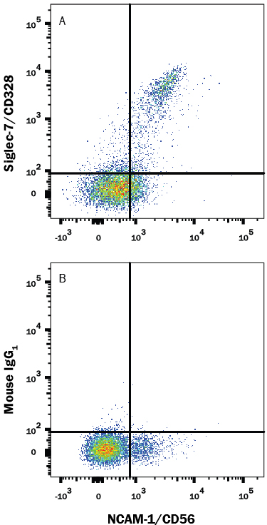
Detection of Siglec‑7/CD328 in Human PBMCs by Flow Cytometry. Human peripheral blood mononuclear cells (PBMCs) were stained with Mouse Anti-Human NCAM-1/CD56 PE-conjugated Monoclonal Antibody (Catalog # FAB2408P) and either (A) Mouse Anti-Human Siglec-7/CD328 Monoclonal Antibody (Catalog # MAB11381) or (B) Mouse IgG1Isotype Control (Catalog # MAB002) followed by Allophycocyanin-conjugated Anti-Mouse IgG Secondary Antibody (Catalog # F0101B). View our protocol for Staining Membrane-associated Proteins.
Reconstitution Calculator
Preparation and Storage
- 12 months from date of receipt, -20 to -70 °C as supplied.
- 1 month, 2 to 8 °C under sterile conditions after reconstitution.
- 6 months, -20 to -70 °C under sterile conditions after reconstitution.
Background: Siglec-7/CD328
Siglecs (1) (sialic acid binding Ig-like lectins) are I-type (Ig-type) lectins belonging to the Ig superfamily. They are characterized by an N-terminal Ig-like V-type domain which mediates sialic acid binding, followed by varying numbers of Ig-like C2-type domains (1, 2). Eleven human Siglecs have been cloned and characterized. They are sialoadhesin/CD169/Siglec-1, CD22/Siglec-2, CD33/Siglec-3, Myelin-Associated Glycoprotein (MAG/Siglec-4a) and Siglecs 5 to 11 (1‑4). To date, no Siglec has been shown to recognized any cell surface ligand other than sialic acids, suggesting that interactions with glycans containing this carbohydrate are important in mediating the biological functions of Siglecs. Siglecs 5 to 11 share a high degree of sequence similarity with CD33/Siglec-3 both in their extracellular and intracellular regions. They are collectively referred to as CD33-related Siglecs. One remarkable feature of the CD33-related Siglecs is their differential expression pattern within the hematopoietic system (2, 3). This fact, together with the presence of two conserved immunoreceptor tyrosine-based inhibition motifs (ITIMs) in their cytoplasmic tails, suggests that CD33-related Siglecs are involved in the regulation of cellular activation within the immune system. The cDNA of human Siglec-7, also known as adhesion inhibitory receptor module-1 (AIRM-1) and designated CD328, encodes a 467 amino acid (aa) polypeptide with a hydrophobic signal peptide, an N-terminal Ig-like V-type domain, two Ig-like C2-type domains, a transmembrane region and a cytoplasmic tail (5). Siglec-7 exists as a monomer on the cell surface and is expressed on natural killer cells, CD8+ T cells and monocytes (3, 5). It binds equally well to both alpha 2,3- and alpha 2,6-linked sialic acid (5). The gene encoding Siglec-7 was mapped to chromosome 19q13.3.
- Crocker, P.R. et al. (1998) Glycobiology 8:v.
- Crocker, P.R. and A. Varki (2001) Trends Immunol. 22:337.
- Crocker, P.R. and A. Varki (2001) Immunology 103:137.
- Angata, T. et al. (2002) J. Biol. Chem. 277:24466.
- Nicoll, G. et al. (1999) J. Biol. Chem. 274:34089.
Product Datasheets
FAQs
No product specific FAQs exist for this product, however you may
View all Antibody FAQsReviews for Human Siglec-7/CD328 Antibody
There are currently no reviews for this product. Be the first to review Human Siglec-7/CD328 Antibody and earn rewards!
Have you used Human Siglec-7/CD328 Antibody?
Submit a review and receive an Amazon gift card.
$25/€18/£15/$25CAN/¥75 Yuan/¥2500 Yen for a review with an image
$10/€7/£6/$10 CAD/¥70 Yuan/¥1110 Yen for a review without an image


