Human SPARC Antibody Summary
Ala18-Ile303
Accession # P09486
Applications
This antibody functions as an ELISA capture antibody when paired with Goat Anti-Human SPARC Antigen Affinity-purified Polyclonal Antibody (Catalog # AF941).
This product is intended for assay development on various assay platforms requiring antibody pairs. We recommend the Human SPARC DuoSet ELISA Kit (Catalog # DY941-05) for convenient development of a sandwich ELISA or the Human SPARC Quantikine ELISA Kit (Catalog # DSP00) for a complete optimized ELISA.
Please Note: Optimal dilutions should be determined by each laboratory for each application. General Protocols are available in the Technical Information section on our website.
Scientific Data
 View Larger
SPARC/Osteonectin in Human Ovary Cancer Tissue. SPARC/Osteonectin was detected in immersion fixed paraffin-embedded sections of human ovarian clear cell carcinoma tissue using Human SPARC/Osteonectin Monoclonal Antibody (Catalog # MAB941) at 25 µg/mL overnight at 4 °C. Tissue was stained using the Anti-Mouse HRP-DAB Cell & Tissue Staining Kit (brown; Catalog # CTS002) and counterstained with hematoxylin (blue). View our protocol for Chromogenic IHC Staining of Paraffin-embedded Tissue Sections.
 View Larger
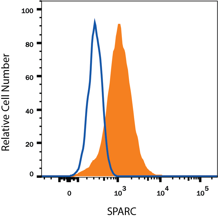
Detection of SPARC in HT1080 Human Cell Line by Flow Cytometry. HT1080 human fibrosarcoma cell line was stained with Mouse Anti-Human SPARC Monoclonal Antibody (Catalog # MAB941, filled histogram) or isotype control antibody (Catalog # MAB002, open histogram), followed by Allophycocyanin-conjugated Anti-Mouse IgG Secondary Antibody (Catalog # F0101B). To facilitate intracellular staining, cells were fixed with Flow Cytometry Fixation Buffer (Catalog # FC004) and permeabilized with Flow Cytometry Permeabilization/Wash Buffer I (Catalog # FC005). View our protocol for Staining Intracellular Molecules.
 View Larger
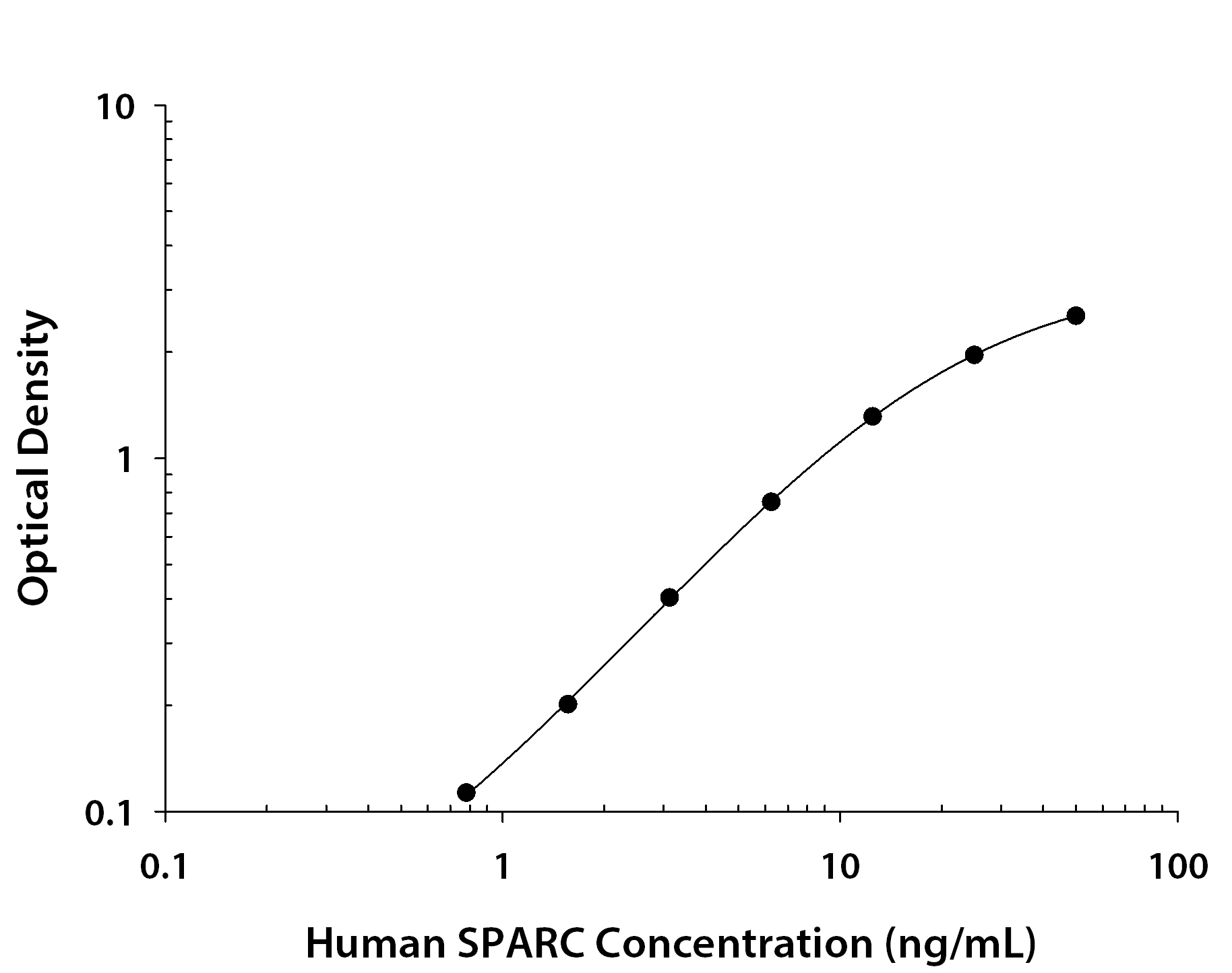
Human SPARC ELISA Standard Curve. Recombinant Human VSIG8 protein was serially diluted 2-fold and captured by Mouse Anti-Human VSIG8 Monoclonal Antibody (Catalog # MAB9418) coated on a Clear Polystyrene Microplate (Catalog # DY990). Goat Anti-Human SPARC Antigen Affinity-purified Polyclonal Antibody (Catalog # AF941) was biotinylated and incubated with the protein captured on the plate. Detection of the standard curve was achieved by incubating Streptavidin-HRP (Catalog # DY998) followed by Substrate Solution (Catalog # DY999) and stopping the enzymatic reaction with Stop Solution (Catalog # DY994).
Reconstitution Calculator
Preparation and Storage
- 12 months from date of receipt, -20 to -70 °C as supplied.
- 1 month, 2 to 8 °C under sterile conditions after reconstitution.
- 6 months, -20 to -70 °C under sterile conditions after reconstitution.
Background: SPARC
SPARC, an acronym for “secreted protein, acidic and rich in cysteine”, is also known as osteonectin or BM-40 (1-5). It is the founding member of a family of secreted matricellular proteins with similar domain structure. The 286 amino acid (aa), 43 kDa protein contains an N-terminal acidic region that binds calcium, a follistatin domain that contains Kazal-like sequences, and a C-terminal extracellular calcium (EC) binding domain with two EF-hand motifs (1-5). Crystal structure modeling shows that residues implicated in cell binding, inhibition of cell spreading, and disassembly of focal adhesions cluster on one face of SPARC, while a collagen binding epitope and an N-glycosylation site are opposite this face (6). SPARC is produced by fibroblasts, capillary endothelial cells, platelets and macrophages, especially in areas of tissue morphogenesis and remodeling (3, 7). SPARC shows context-specific effects, but generally inhibits adhesion, spreading and proliferation, and promotes collagen matrix formation (3-5). For endothelial cells, SPARC disrupts focal adhesions and binds and sequesters PDGF and VEGF (3-5). SPARC is abundantly expressed in bone, where it promotes osteoblast differentiation and inhibits adipogenesis (5, 8). SPARC is potentially cleaved by metalloproteinases, producing an angiogenic peptide that includes the copper-binding sequence KGHK (7). Paradoxically, SPARC is highly expressed in many tumor types undergoing an endothelial to mesenchymal transition; its expression, however, mainly decreases the likelihood of metastasis and confers sensitivity to chemotherapy and radiation (4, 9-11). Stabilin-1, which is expressed on alternately activated macrophages, is the first SPARC receptor to be identified. It binds the SPARC EC domain and mediates endocytosis for degradation (12). Mature human SPARC shows 92%, 92%, 97%, 99%, 96% and 85% aa identity with mouse, rat, canine, bovine, porcine and chick SPARC, respectively.
- Lankat-Buttgereit, B. et al. (1988) FEBS Lett. 236:352.
- Sweetwyne, M. T. et al. (2004) J. Histochem. Cytochem. 52:723.
- Sage, H. et al. (1989) J. Cell Biol. 109:341.
- Framson, P. E. and E. H. Sage (2004) J. Cell. Biochem. 92:679.
- Alford, A. I. and K. D. Hankenson (2006) Bone 38:749.
- Hohenester, E et al. (1997) EMBO J. 16:3778.
- Sage, E. H. et al. (2003) J. Biol. Chem. 278:37849.
- Delany, A. M. et al. (2003) Endocrinology 144:2588.
- Robert, G. et al. (2006) Cancer Res. 66:7516.
- Koblinski, J. E. et al. (2005) Cancer Res. 65:7370.
- Tai, I. T. et al. (2005) J. Clin. Invest. 115:1492.
- Kzhyshkowska, J. et al. (2006) J. Immunol. 176:5825.
Product Datasheets
Citations for Human SPARC Antibody
R&D Systems personnel manually curate a database that contains references using R&D Systems products. The data collected includes not only links to publications in PubMed, but also provides information about sample types, species, and experimental conditions.
5
Citations: Showing 1 - 5
Filter your results:
Filter by:
-
Osteoblast differentiation of equine induced pluripotent stem cells
Authors: A Baird, T Lindsay, A Everett, V Iyemere, YZ Paterson, A McClellan, FMD Henson, DJ Guest
Biol Open, 2018-05-10;0(0):.
Species: Equine
Sample Types: Whole Cells
Applications: ICC -
Summary of expression of SPARC protein in cutaneous vascular neoplasms and mimickers
Authors: SH Mauzo, DR Milton, VG Prieto, CA Torres-Cab, WL Wang, N Chakravart, P Nagarajan, MT Tetzlaff, JL Curry, D Ivan, RE Brown, PP Aung
Ann Diagn Pathol, 2018-03-15;34(0):151-154.
Species: Human
Sample Types: Whole Tissue
Applications: IHC-P -
Potential actionable targets in appendiceal cancer detected by immunohistochemistry, fluorescent in situ hybridization, and mutational analysis
Authors: E Borazanci, SZ Millis, J Kimbrough, N Doll, D Von Hoff, RK Ramanathan
J Gastrointest Oncol, 2017-02-01;8(1):164-172.
Species: Human
Sample Types: Whole Tissue
Applications: IHC-P -
Insulin-like growth factor binding protein-4 (IGFBP-4) is a novel anti-angiogenic and anti-tumorigenic mediator secreted by dibutyryl cyclic AMP (dB-cAMP)-differentiated glioblastoma cells.
Authors: Moreno MJ, Ball M, Andrade MF, McDermid A, Stanimirovic DB
Glia, 2006-06-01;53(8):845-57.
Species: Human
Sample Types: Cell Culture Supernates
Applications: ELISA Development -
Biomarker discovery from pancreatic cancer secretome using a differential proteomic approach.
Authors: Gronborg M, Kristiansen TZ, Iwahori A, Chang R, Reddy R, Sato N, Molina H, Jensen ON, Hruban RH, Goggins MG, Maitra A, Pandey A
Mol. Cell Proteomics, 2005-10-08;5(1):157-71.
Species: Human
Sample Types: Cell Lysates
Applications: Western Blot
FAQs
No product specific FAQs exist for this product, however you may
View all Antibody FAQsReviews for Human SPARC Antibody
Average Rating: 5 (Based on 2 Reviews)
Have you used Human SPARC Antibody?
Submit a review and receive an Amazon gift card.
$25/€18/£15/$25CAN/¥75 Yuan/¥2500 Yen for a review with an image
$10/€7/£6/$10 CAD/¥70 Yuan/¥1110 Yen for a review without an image
Filter by:
This antibody was used as capture with AF941 as the detection. It worked great in an ELISA to measure human serum and plasma samples.


