Mouse BTLA Antibody

Catalog # Availability Size / Price Qty
AF3007
AF3007-SP
Detection of Mouse BTLA by Western Blot.
6 Images
Product Details
Citations (2)
FAQs
Supplemental Products
Reviews

Mouse BTLA Antibody Summary

Species Reactivity
Mouse
Specificity
Detects mouse BTLA in direct ELISAs and Western blots. In direct ELISAs and Western blots, less than 1% cross-reactivity with recombinant human BTLA is observed.
Source
Polyclonal Goat IgG
Purification
Antigen Affinity-purified
Immunogen
Mouse myeloma cell line NS0-derived recombinant mouse BTLA
Glu30-Gly176 (Pro41Glu, Thr45Asn, Thr47Lys, Gln52His, Arg55Trp, Gln63Glu, Cys85Trp)
Accession # Q32MV9
Formulation
Lyophilized from a 0.2 μm filtered solution in PBS with Trehalose. *Small pack size (SP) is supplied either lyophilized or as a 0.2 µm filtered solution in PBS.

Applications

Recommended Concentration
Sample
Western Blot
1 µg/mL
See below

Please Note: Optimal dilutions should be determined by each laboratory for each application. General Protocols are available in the Technical Information section on our website.

Scientific Data
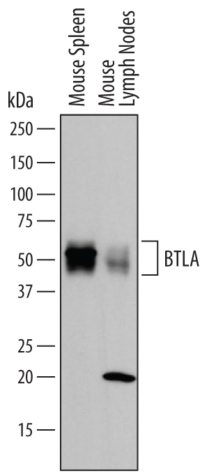
Western Blot Detection of Mouse BTLA antibody by Western Blot. View Larger

Detection of Mouse BTLA by Western Blot. Western blot shows lysates of mouse spleen tissue and mouse lymph node tissue. PVDF membrane was probed with 1 µg/mL of Goat Anti-Mouse BTLA Antigen Affinity-purified Polyclonal Antibody (Catalog # AF3007) followed by HRP-conjugated Anti-Goat IgG Secondary Antibody (Catalog # HAF109). A specific band was detected for BTLA at approximately 60 kDa (as indicated). This experiment was conducted under reducing conditions and using Immunoblot Buffer Group 1.

Western Blot Detection of Human BTLA/CD272 by Western Blot View Larger

Detection of Human BTLA/CD272 by Western Blot Activation of PPAR beta /δ results in upregulation of FABP5. (a) PC3M cells were treated with GW0742 (1 μM, 4 h). Expression of FABP5, PDK1, ADRP, and VEGF were assessed by Q-PCR. The data were normalized to the untreated control. (b, c) Denoted cells were treated with 1 μM (a) or 2 μM (b) GW0742 for 16 h. Left panels, immunoblots of FABP5 in untreated versus GW0742-treated cells. Right panel: Densitometry analyses depicting changes in FABP5 expression upon treatment with GW0742 in three independent experiments (mean ± S.D.). (d, e) PC3M cells were transfected with vectors harboring SiScramble or PPAR beta /δ siRNA. Three days later, expression of FABP5, PPAR beta /δ, and VEGF mRNA in were measured by Q-PCR (d) and FABP5 expression was assessed by immunoblot (e). (f) PC3M cells were stably transfected with an empty vector (PGIPZ), or a vector harbouring shFABP5. Expression levels of FABP5, PPAR beta /δ, and VEGF mRNA were measured by Q-PCR. *P <.05 versus nontreated controls. Image collected and cropped by CiteAb from the following publication (https://pubmed.ncbi.nlm.nih.gov/20847935), licensed under a CC-BY license. Not internally tested by R&D Systems.

Western Blot Detection of Human BTLA/CD272 by Western Blot View Larger

Detection of Human BTLA/CD272 by Western Blot Prostate cancer progression is accompanied by up regulation of FABP5/PPAR beta /δ expression and signalling. (a) Bottom: Expression levels of FABP5 mRNA in denoted cell lines were measured by Q-PCR. Top: Level of FABP5 protein in denoted cell lines assessed by immunoblots. (b) Bottom: Expression levels of PDK-1 and PPAR beta /δ mRNA in denoted cell lines were measured by Q-PCR. Top: Immunoblots of PDK1 in denoted cell lines. Data are mean ± S.D. (n = 3). *P <.02 versus 22Rv.1. (Paired T test). Immunoblots were repeated three times with similar results. Image collected and cropped by CiteAb from the following publication (https://pubmed.ncbi.nlm.nih.gov/20847935), licensed under a CC-BY license. Not internally tested by R&D Systems.

Western Blot Detection of Human BTLA/CD272 by Western Blot View Larger

Detection of Human BTLA/CD272 by Western Blot Activation of PPAR beta /δ results in upregulation of FABP5. (a) PC3M cells were treated with GW0742 (1 μM, 4 h). Expression of FABP5, PDK1, ADRP, and VEGF were assessed by Q-PCR. The data were normalized to the untreated control. (b, c) Denoted cells were treated with 1 μM (a) or 2 μM (b) GW0742 for 16 h. Left panels, immunoblots of FABP5 in untreated versus GW0742-treated cells. Right panel: Densitometry analyses depicting changes in FABP5 expression upon treatment with GW0742 in three independent experiments (mean ± S.D.). (d, e) PC3M cells were transfected with vectors harboring SiScramble or PPAR beta /δ siRNA. Three days later, expression of FABP5, PPAR beta /δ, and VEGF mRNA in were measured by Q-PCR (d) and FABP5 expression was assessed by immunoblot (e). (f) PC3M cells were stably transfected with an empty vector (PGIPZ), or a vector harbouring shFABP5. Expression levels of FABP5, PPAR beta /δ, and VEGF mRNA were measured by Q-PCR. *P <.05 versus nontreated controls. Image collected and cropped by CiteAb from the following publication (https://pubmed.ncbi.nlm.nih.gov/20847935), licensed under a CC-BY license. Not internally tested by R&D Systems.

Western Blot Detection of Human BTLA/CD272 by Western Blot View Larger

Detection of Human BTLA/CD272 by Western Blot The FABP5/PPAR beta /δ pathway enhances prostate cancer cell proliferation and transformation. (a) PC3M cells that stably express shFABP5 were cultured in a 24 well plate (2500 cells/well) and treated with vehicle or GW 0742 (1 μM). Cells were counted at the denoted days. Data are mean ± SEM (n = 3). Inset: immunoblotting demonstrating low FABP5 level in shFABP5-expressing. (b) PC3M cells were transfected with a vector harboring PPAR beta /δsiRNA. Four days later, were cultured in a 24 well plate (2500 cells/well) and treated with vehicle or GW 0742 (1 μM). Cells were counted at the denoted days. Inset: Q-PCR analyses demonstrating low PPAR beta /δ in cells transfected with SiRNA towards the receptor. (c) Colony formation assays were conducted in the absence or presence of GW0742 (1 μM). Cells were cultured in 2% agarose for 29 days (see Materials and methods for details). Media was replenished every 4 days. Colonies were visualized by staining with 0.005% crystal violet and counted under a light microscope. Data are mean ± SEM (n = 3). Image collected and cropped by CiteAb from the following publication (https://pubmed.ncbi.nlm.nih.gov/20847935), licensed under a CC-BY license. Not internally tested by R&D Systems.

Western Blot Detection of Human BTLA/CD272 by Western Blot View Larger

Detection of Human BTLA/CD272 by Western Blot Activation of PPAR beta /δ results in upregulation of FABP5. (a) PC3M cells were treated with GW0742 (1 μM, 4 h). Expression of FABP5, PDK1, ADRP, and VEGF were assessed by Q-PCR. The data were normalized to the untreated control. (b, c) Denoted cells were treated with 1 μM (a) or 2 μM (b) GW0742 for 16 h. Left panels, immunoblots of FABP5 in untreated versus GW0742-treated cells. Right panel: Densitometry analyses depicting changes in FABP5 expression upon treatment with GW0742 in three independent experiments (mean ± S.D.). (d, e) PC3M cells were transfected with vectors harboring SiScramble or PPAR beta /δ siRNA. Three days later, expression of FABP5, PPAR beta /δ, and VEGF mRNA in were measured by Q-PCR (d) and FABP5 expression was assessed by immunoblot (e). (f) PC3M cells were stably transfected with an empty vector (PGIPZ), or a vector harbouring shFABP5. Expression levels of FABP5, PPAR beta /δ, and VEGF mRNA were measured by Q-PCR. *P <.05 versus nontreated controls. Image collected and cropped by CiteAb from the following publication (https://pubmed.ncbi.nlm.nih.gov/20847935), licensed under a CC-BY license. Not internally tested by R&D Systems.

Reconstitution Calculator

Reconstitution Calculator

The reconstitution calculator allows you to quickly calculate the volume of a reagent to reconstitute your vial. Simply enter the mass of reagent and the target concentration and the calculator will determine the rest.

=
÷

Preparation and Storage

Reconstitution
Reconstitute at 0.2 mg/mL in sterile PBS.
Loading...
Shipping
Lyophilized product is shipped at ambient temperature. Liquid small pack size (-SP) is shipped with polar packs. Upon receipt, store immediately at the temperature recommended below.
Stability & Storage
Use a manual defrost freezer and avoid repeated freeze-thaw cycles.
  • 12 months from date of receipt, -20 to -70 °C as supplied.
  • 1 month, 2 to 8 °C under sterile conditions after reconstitution.
  • 6 months, -20 to -70 °C under sterile conditions after reconstitution.

Background: BTLA

B- and T-lymphocyte attenuator (BTLA; CD272) is a 70 kDa, Ig-superfamily, type I transmembrane glycoprotein that is structurally similar to the CD28 family of T cell co-stimulatory or coinhibitory molecules (1‑3). Unlike CD28 family members, however, the BTLA extracellular Ig domain is an I-type rather than a V-type domain, and BTLA does not form homodimers (4). BTLA also differs from CD28 family members through the interaction of its Ig domain with the TNF superfamily member HVEM (herpesvirus entry mediator; TNFSF14) rather than with B7 family ligands (5). BTLA is a coinhibitory molecule expressed on T cells, B cells and, depending on the mouse strain, macrophages, dendritic and NK cells (6). Expression is low in naïve T cells and increased during antigen-specific induction of anergy. In B cells, BTLA is highest when cells are mature and naïve (6). BTLA apparently limits T cell numbers, since deletion of BTLA results in overproduction of T cells, especially CD8+ memory T cells that are hyper-responsive to TCR crosslinking (7). The 305 amino acid (aa) BTLA contains a 29 aa signal sequence, a 153 aa extracellular domain (ECD), a 21 aa transmembrane sequence, and a 102 aa cytoplasmic domain. There are two ITIM motifs and three Tyr phosphorylation sites in the cytoplasmic tail that mediate inhibitory signaling (8, 9). The binding of the BTLA to HVEM does not preclude additional binding of a mammalian stimulatory HVEM ligand, either LIGHT or lymphotoxin-alpha to the complex (4). At least three alleles varying by up to ten extracellular amino acids occur in different mouse strains (6). The ECD of C57BL/6 BTLA shows 51%, 77% and 40% aa identity to that of human, rat and canine BTLA, respectively. A splice variant lacking the Ig domain, termed BTLAs, has been reported (3).

References
  1. Murphy, K. M. et al. (2006) Nat. Rev. Immunol. 6:671.
  2. Croft, M. (2005) Trends Immunol. 26:292.
  3. Watanabe, N. et al. (2003) Nat. Immunol. 4:670.
  4. Compaan, D. M. et al. (2005) J. Biol. Chem. 280:39553.
  5. Sedy, J. R. et al. (2005) Nat. Immunol. 6:90.
  6. Hurchla, M. A. et al. (2005) J. Immunol. 174:3377.
  7. Krieg, C. et al. (2007) Nat. Immunol. 8:162.
  8. Gavrieli, M. et al. (2003) Biochem. Biophys. Res. Commun. 312:1236.
  9. Chemnitz, J. M. et al. (2006) J. Immunol. 176:6603.
Long Name
B- And T-Lymphocyte Attenuator
Entrez Gene IDs
151888 (Human); 208154 (Mouse); 102144877 (Cynomolgus Monkey)
Alternate Names
B and T lymphocyte associated; B and T lymphocyte attenuator; B- and T-lymphocyte attenuator; B- and T-lymphocyte-associated protein; BTLA; BTLA1; CD272 antigen; CD272; FLJ16065; MGC129743

Product Datasheets

You must select a language.

x

Citations for Mouse BTLA Antibody

R&D Systems personnel manually curate a database that contains references using R&D Systems products. The data collected includes not only links to publications in PubMed, but also provides information about sample types, species, and experimental conditions.

2 Citations: Showing 1 - 2
Filter your results:

Filter by:

  1. Quantitative Interactomics in Primary T Cells Provides a Rationale for Concomitant PD-1 and BTLA Coinhibitor Blockade in Cancer Immunotherapy
    Authors: J Celis-Guti, P Blattmann, Y Zhai, N Jarmuzynsk, K Ruminski, C Grégoire, Y Ounoughene, F Fiore, R Aebersold, R Roncagalli, M Gstaiger, B Malissen
    Cell Rep, 2019-06-11;27(11):3315-3330.e7.
    Species: Mouse
    Sample Types: Cell Lysates
    Applications: Western Blot
  2. Involvement of Fatty Acid Binding Protein 5 and PPAR beta /δ in Prostate Cancer Cell Growth
    Authors: Elwin Morgan, Padmamalini Kannan-Thulasiraman, Noa Noy
    PPAR Research

FAQs

No product specific FAQs exist for this product, however you may

View all Antibody FAQs
Loading...

Reviews for Mouse BTLA Antibody

There are currently no reviews for this product. Be the first to review Mouse BTLA Antibody and earn rewards!

Have you used Mouse BTLA Antibody?

Submit a review and receive an Amazon gift card.

$25/€18/£15/$25CAN/¥75 Yuan/¥2500 Yen for a review with an image

$10/€7/£6/$10 CAD/¥70 Yuan/¥1110 Yen for a review without an image

Submit a Review