Mouse Cathepsin E Antibody Summary
Applications
Please Note: Optimal dilutions should be determined by each laboratory for each application. General Protocols are available in the Technical Information section on our website.
Scientific Data
 View Larger
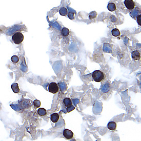
Cathepsin E in Mouse Lung. Cathepsin E was detected in perfusion fixed frozen sections of mouse lung using Goat Anti-Mouse Cathepsin E Antigen Affinity-purified Polyclonal Antibody (Catalog # AF1130) at 15 µg/mL overnight at 4 °C. Tissue was stained using the Anti-Goat HRP-DAB Cell & Tissue Staining Kit (brown; Catalog # CTS008) and counterstained with hematoxylin (blue). Specific labeling was localized to the plasma membrane of type II alveolar cells. View our protocol for Chromogenic IHC Staining of Frozen Tissue Sections.
Reconstitution Calculator
Preparation and Storage
- 12 months from date of receipt, -20 to -70 degreesC as supplied. 1 month, 2 to 8 degreesC under sterile conditions after reconstitution. 6 months, -20 to -70 degreesC under sterile conditions after reconstitution.
Background: Cathepsin E
Cathepsin E is an intracellular aspartic protease of the pepsin family (1, 2). Unlike Cathepsin D, another member of the same family and a lysosomal protease with relatively ubiquitous distribution, Cathepsin E is not a lysosomal enzyme and has a limited cell and tissue distribution. However, both Cathepsins D and E play an important role in the degradation of proteins, the generation of bioactive proteins, and antigen processing (3). Both enzymes are efficient in cleaving the Swedish mutant of amyloid precursor protein (APP) at the beta site but show almost no reactivity with the wild-type APP (4). Mouse Cathepsin E is synthesized as a precursor protein, consisting of a signal peptide (residues 1-18), a propeptide (residues 19-59), and a mature chain (residues 60-397) (1).
- Tatnell, P.J. et al. (1997) FEBS Lett. 408:62.
- Kay, J. and P.J. Tatnell (2004) in Handbook of Proteolytic Enzymes (Barrett, A.J. et al., eds.), p. 33, Academic Press, San Diego.
- Tsukuba, T. et al. (2000) Mol. Cells 10:601.
- Gruninger-Leitch, F. et al. (2000) Nat. Biotechnol. 18:66.
Product Datasheets
Citations for Mouse Cathepsin E Antibody
R&D Systems personnel manually curate a database that contains references using R&D Systems products. The data collected includes not only links to publications in PubMed, but also provides information about sample types, species, and experimental conditions.
2
Citations: Showing 1 - 2
Filter your results:
Filter by:
-
Global protease activity profiling provides differential diagnosis of pancreatic cysts
Authors: SL Ivry, JM Sharib, DA Dominguez, N Roy, SE Hatcher, M Yip-Schnei, CM Schmidt, RE Brand, WG Park, M Hebrok, G Kim, AJ O'Donoghue, KS Kirkwood, CS Craik
Clin. Cancer Res., 2017-04-19;0(0):.
Species: Mouse
Sample Types: Whole Tissue
Applications: IHC-P -
Gene targeting of the cysteine peptidase cathepsin H impairs lung surfactant in mice.
Authors: Buhling F, Kouadio M, Chwieralski CE, Kern U, Hohlfeld JM, Klemm N, Friedrichs N, Roth W, Deussing JM, Peters C, Reinheckel T
PLoS ONE, 2011-10-12;6(10):e26247.
Species: Mouse
Sample Types: Tissue Homogenates
Applications: Western Blot
FAQs
No product specific FAQs exist for this product, however you may
View all Antibody FAQsReviews for Mouse Cathepsin E Antibody
Average Rating: 5 (Based on 1 Review)
Have you used Mouse Cathepsin E Antibody?
Submit a review and receive an Amazon gift card.
$25/€18/£15/$25CAN/¥75 Yuan/¥2500 Yen for a review with an image
$10/€7/£6/$10 CAD/¥70 Yuan/¥1110 Yen for a review without an image
Filter by:


