Mouse CCL3/MIP-1 alpha Antibody Summary
Applications
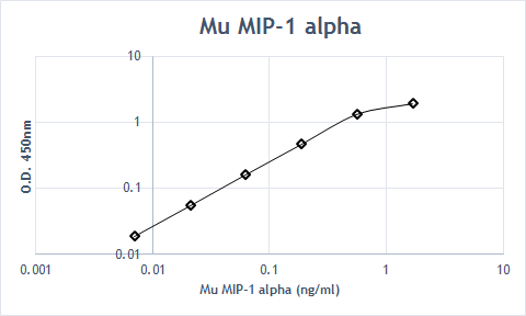
Mouse CCL3/MIP-1 alpha Sandwich Immunoassay
Please Note: Optimal dilutions should be determined by each laboratory for each application. General Protocols are available in the Technical Information section on our website.
Scientific Data
 View Larger
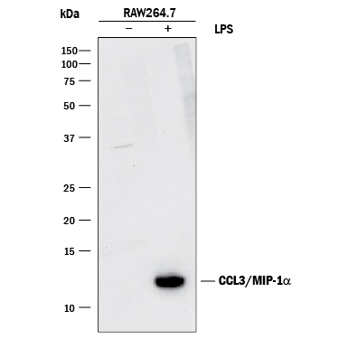
Detection of Mouse CCL3/MIP‑1 alpha by Western Blot. Western blot shows lysates of RAW 264.7 mouse monocyte/macrophage cell line untreated (-) or treated (+) with 10 µg/mL LPS for 4 hours. PVDF membrane was probed with 1 µg/mL of Goat Anti-Mouse CCL3/MIP-1a Antigen Affinity-purified Polyclonal Antibody (Catalog # AF-450-NA) followed by HRP-conjugated Anti-Goat IgG Secondary Antibody (Catalog # HAF017). A specific band was detected for CCL3/MIP-1a at approximately 12 kDa (as indicated). This experiment was conducted under reducing conditions and using Immunoblot Buffer Group 1.
 View Larger
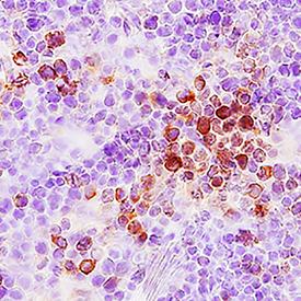
CCL3/MIP‑1 alpha in Mouse Thymus. CCL3/MIP-1a was detected in perfusion fixed frozen sections of mouse thymus using Goat Anti-Mouse CCL3/MIP-1a Antigen Affinity-purified Polyclonal Antibody (Catalog # AF-450-NA) at 15 µg/mL overnight at 4 °C. Tissue was stained using the Anti-Goat HRP-DAB Cell & Tissue Staining Kit (brown; Catalog # CTS008) and counterstained with hematoxylin (blue). Specific staining was localized to cytoplasm. View our protocol for Chromogenic IHC Staining of Frozen Tissue Sections.
 View Larger
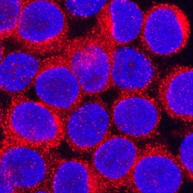
CCL3/MIP‑1 alpha in RAW 264.7 Mouse Cell Line. CCL3/MIP-1a was detected in immersion fixed RAW 264.7 mouse monocyte/macrophage cell line using Goat Anti-Mouse CCL3/MIP-1a Antigen Affinity-purified Polyclonal Antibody (Catalog # AF-450-NA) at 5 µg/mL for 3 hours at room temperature. Cells were stained using the NorthernLights™ 557-conjugated Anti-Goat IgG Secondary Antibody (red; Catalog # NL001) and counterstained with DAPI (blue). Specific staining was localized to cytoplasm. View our protocol for Fluorescent ICC Staining of Cells on Coverslips.
 View Larger
Chemotaxis Induced by CCL3/MIP‑1 alpha and Neutralization by Mouse CCL3/MIP‑1 alpha Antibody. Recombinant Mouse CCL3/MIP-1a (Catalog # 450-MA) chemoattracts the BaF3 mouse pro-B cell line transfected with human CCR5 in a dose-dependent manner (orange line). The amount of cells that migrated through to the lower chemotaxis chamber was measured by Resazurin (Catalog # AR002). Chemotaxis elicited by Recombinant Mouse CCL3/ MIP-1a (10 ng/mL) is neutralized (green line) by increasing concentrations of Goat Anti-Mouse CCL3/MIP-1a Antigen Affinity-purified Polyclonal Antibody (Catalog # AF-450-NA). The ND50 is typically 0.05-0.2 µg/mL.
Reconstitution Calculator
Preparation and Storage
- 12 months from date of receipt, -20 to -70 degreesC as supplied. 1 month, 2 to 8 degreesC under sterile conditions after reconstitution. 6 months, -20 to -70 degreesC under sterile conditions after reconstitution.
Background: CCL3/MIP-1 alpha
The macrophage inflammatory proteins 1 alpha and 1 beta, two closely related but distinct proteins, were originally co-purified from medium conditioned by a LPS-stimulated murine macrophage cell line. Mature mouse MIP-1 alpha shares approximately 77% and 70% amino acid identity with human MIP-1 alpha and mouse MIP-1 beta, respectively. MIP-1 proteins are expressed primarily in T cells, B cells, and monocytes after antigen or mitogen stimulation. The MIP-1 proteins are members of the beta (C-C) subfamily of chemokines. Both MIP-1 alpha and MIP-1 beta are monocyte chemoattractants in vitro. Additionally, the MIP-1 proteins have been reported to have chemoattractant and adhesive effects on lymphocytes, with MIP-1 alpha and MIP-1 beta preferentially attracting CD8 + and CD4 + T cells, respectively. MIP-1 alpha has also been shown to attract B cells as well as eosinophils. MIP-1 proteins have been reported to have multiple effects on hematopoietic precursor cells and MIP-1 alpha has been identified as a stem cell inhibitory factor that can inhibit the proliferation of hematopoietic stem cells in vitro as well as in vivo. In the same assays, MIP-1 beta was reported to be much less active. The functional receptor for MIP-1 alpha has been identified as CCR1 and CCR5.
Product Datasheets
Citations for Mouse CCL3/MIP-1 alpha Antibody
R&D Systems personnel manually curate a database that contains references using R&D Systems products. The data collected includes not only links to publications in PubMed, but also provides information about sample types, species, and experimental conditions.
20
Citations: Showing 1 - 10
Filter your results:
Filter by:
-
Deficiency of lncRNA MERRICAL abrogates macrophage chemotaxis and diabetes-associated atherosclerosis
Authors: Chen, J;Jamaiyar, A;Wu, W;Hu, Y;Zhuang, R;Sausen, G;Cheng, HS;de Oliveira Vaz, C;Pérez-Cremades, D;Tzani, A;McCoy, MG;Assa, C;Eley, S;Randhawa, V;Lee, K;Plutzky, J;Hamburg, NM;Sabatine, MS;Feinberg, MW;
Cell reports
Species: Murine polyomavirus strain A3
Sample Types: Whole Cells
Applications: Neutralization -
Neutrophil-dendritic cell interaction plays an important role in live attenuated Leishmania vaccine induced immunity
Authors: P Bhattachar, N Ismail, A Saxena, S Gannavaram, R Dey, T Oljuskin, A Akue, K Takeda, J Yu, S Karmakar, PK Dagur, JP McCoy, HL Nakhasi
PloS Neglected Tropical Diseases, 2022-02-22;16(2):e0010224.
Species: Mouse
Sample Types: In Vivo
Applications: In Vivo -
Radiation Triggers a Dynamic Sequence of Transient Microglial Alterations in Juvenile Brain
Authors: AM Osman, Y Sun, TC Burns, L He, N Kee, N Oliva-Vila, A Alevyzaki, K Zhou, L Louhivuori, P Uhlén, E Hedlund, C Betsholtz, VM Lauschke, J Kele, K Blomgren
Cell Rep, 2020-06-02;31(9):107699.
Species: Mouse
Sample Types:
Applications: IHC -
Regulation of the bi-directional cross-talk between ovarian cancer cells and adipocytes by SPARC
Authors: B John, C Naczki, C Patel, A Ghoneum, S Qasem, Z Salih, N Said
Oncogene, 2019-02-14;0(0):.
Species: Mouse
Sample Types: In Vivo
Applications: Neutralization -
B cells inhibit bone formation in rheumatoid arthritis by suppressing osteoblast differentiation
Authors: W Sun, N Meednu, A Rosenberg, J Rangel-Mor, V Wang, J Glanzman, T Owen, X Zhou, H Zhang, BF Boyce, JH Anolik, L Xing
Nat Commun, 2018-12-03;9(1):5127.
Species: Mouse
Sample Types: Whole Cells
Applications: Neutralization -
IFN? Protects Neurons from Damage in a Murine Model of HIV-1 Associated Brain Injury
Authors: VE Thaney, AM O'Neill, MM Hoefer, R Maung, AB Sanchez, M Kaul
Sci Rep, 2017-04-20;7(0):46514.
Species: Mouse, Rat
Sample Types: Whole Cells
Applications: Neutralization -
Hippocampal T cell infiltration promotes neuroinflammation and cognitive decline in a mouse model of tauopathy
Authors: David Blum
Brain, 2016-11-05;0(0):.
Species: Mouse
Sample Types: Whole Tissue
Applications: IHC -
Response patterns of cytokines/chemokines in two murine strains after irradiation.
Authors: Zhang M, Yin L, Zhang K, Sun W, Yang S, Zhang B, Salzman P, Wang W, Liu C, Vidyasagar S, Zhang L, Ju S, Okunieff P, Zhang L
Cytokine, 2012-01-25;58(2):169-77.
Species: Mouse
Sample Types: Plasma
Applications: Luminex Development -
Diesel exhaust particulates exacerbate asthma-like inflammation by increasing CXC chemokines.
Authors: Kim J, Natarajan S, Vaickus LJ, Bouchard JC, Beal D, Cruikshank WW, Remick DG
Am. J. Pathol., 2011-10-01;179(6):2730-9.
Species: Mouse
Sample Types: BALF
Applications: ELISA Development -
Comprehensive assessment of chemokine expression profiles by flow cytometry.
Authors: Eberlein J, Nguyen TT, Victorino F, Golden-Mason L, Rosen HR, Homann D
J. Clin. Invest., 2010-02-08;120(3):907-23.
Species: Mouse
Sample Types: Whole Cells
Applications: Flow Cytometry -
Recruitment and differentiation of conventional dendritic cell precursors in tumors.
Authors: Diao J, Zhao J, Winter E, Cattral MS
J. Immunol., 2009-12-21;184(3):1261-7.
Species: Mouse
Sample Types: In Vivo
Applications: Neutralization -
Resistance of human alveolar macrophages to Bacillus anthracis lethal toxin.
Authors: Wu W, Mehta H, Chakrabarty K, Booth JL, Duggan ES, Patel KB, Ballard JD, Coggeshall KM, Metcalf JP
J. Immunol., 2009-10-07;183(9):5799-806.
Species: Mouse
Sample Types: Cell Culture Supernates
Applications: ELISA Development -
Increased cytokine production in IL-18 receptor alpha-deficient cells is associated with dysregulation of suppressors of cytokine signaling (SOCS).
Authors: Nold-Petry CA, Nold MF, Nielsen JW, Bustamante A, Zepp JA, Storm KA, Hong JW, Kim SH, Dinarello CA
J. Biol. Chem., 2009-07-10;0(0):.
Species: Mouse
Sample Types: Cell Culture Supernates
Applications: ELISA Development -
CCL3-CCR5 axis regulates intratumoral accumulation of leukocytes and fibroblasts and promotes angiogenesis in murine lung metastasis process.
Authors: Wu Y, Li YY, Matsushima K, Baba T, Mukaida N
J. Immunol., 2008-11-01;181(9):6384-93.
Species: Mouse
Sample Types: Whole Tissue
Applications: IHC-P -
Role of the chemokine decoy receptor D6 in balancing inflammation, immune activation, and antimicrobial resistance in Mycobacterium tuberculosis infection.
Authors: Di Liberto D, Locati M, Caccamo N, Vecchi A, Meraviglia S, Salerno A, Sireci G, Nebuloni M, Caceres N, Cardona PJ, Dieli F, Mantovani A
J. Exp. Med., 2008-08-11;205(9):2075-84.
Species: Mouse
Sample Types: In Vivo
Applications: Neutralization -
Neutrophil recruitment in immunized mice depends on MIP-2 inducing the sequential release of MIP-1alpha, TNF-alpha and LTB(4).
Authors: Ramos CD, Fernandes KS, Canetti C, Teixeira MM, Silva JS, Cunha FQ
Eur. J. Immunol., 2006-08-01;36(8):2025-34.
Species: Mouse
Sample Types: Tissue Secretion
Applications: ELISA Development -
Selective macrophage suppression during sepsis.
Authors: Ellaban E, Bolgos G, Remick D
Cell. Immunol., 2005-02-26;231(1):103-11.
Species: Mouse
Sample Types: Cell Culture Supernates
Applications: ELISA Development -
Involvement of monocyte chemoattractant protein-1, macrophage inflammatory protein-1alpha and interleukin-1beta in Wallerian degeneration.
Authors: Perrin FE, Lacroix S, Aviles-Trigueros M, David S
Brain, 2005-02-02;128(0):854-66.
Species: Mouse
Sample Types: In Vivo
Applications: Neutralization -
CC chemokines mediate leukocyte trafficking into the central nervous system during murine neurocysticercosis: role of gamma delta T cells in amplification of the host immune response.
Authors: Cardona AE, Gonzalez PA, Teale JM
Infect. Immun., 2003-05-01;71(5):2634-42.
Species: Mouse
Sample Types: Whole Cells
Applications: ICC -
Immunostimulatory DNA-based vaccines elicit multifaceted immune responses against HIV at systemic and mucosal sites.
Authors: Horner AA, Datta SK, Takabayashi K, Belyakov IM, Cinman N, Nguyen MD, Van Uden JH, Berzofsky JA
J. Immunol., 2001-08-01;167(3):1584-91.
Species: Mouse
Sample Types: Whole Cells
Applications: ELISpot Development
FAQs
No product specific FAQs exist for this product, however you may
View all Antibody FAQsReviews for Mouse CCL3/MIP-1 alpha Antibody
Average Rating: 4.3 (Based on 3 Reviews)
Have you used Mouse CCL3/MIP-1 alpha Antibody?
Submit a review and receive an Amazon gift card.
$25/€18/£15/$25CAN/¥75 Yuan/¥2500 Yen for a review with an image
$10/€7/£6/$10 CAD/¥70 Yuan/¥1110 Yen for a review without an image
Filter by:


