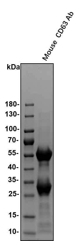
Mouse CD63 Antibody Summary
Met1-Met238
Accession # P41731
Applications
Please Note: Optimal dilutions should be determined by each laboratory for each application. General Protocols are available in the Technical Information section on our website.
Scientific Data
 View Larger
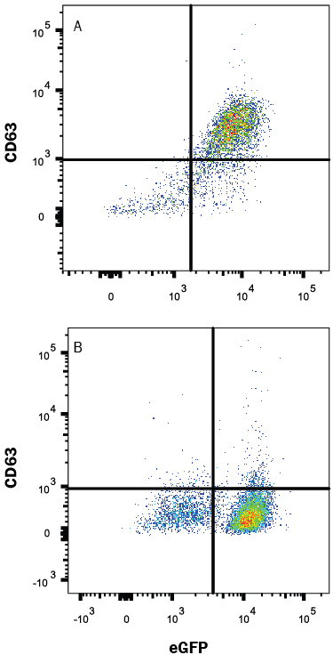
Detection of CD63 in HEK293 Human Cell Line Transfected with Mouse CD63 and eGFP by Flow Cytometry. HEK293 human embryonic kidney cell line transfected with either (A) mouse CD63 or (B) irrelevant transfectants and eGFP were stained with Rat Anti-Mouse CD63 Monoclonal Antibody (Catalog # MAB5417) followed by Allophycocyanin-conjugated Anti-Rat IgG Secondary Antibody (Catalog # F0113). Quadrant markers were set based on control antibody staining (Catalog # MAB005). View our protocol for Staining Membrane-associated Proteins.
 View Larger
CD63 in Mouse Kidney. CD63 was detected in perfusion fixed frozen sections of mouse kidney using Rat Anti-Mouse CD63 Monoclonal Antibody (Catalog # MAB5417) at 25 µg/mL overnight at 4 °C. Tissue was stained using the Anti-Rat HRP-DAB Cell & Tissue Staining Kit (brown; Catalog # CTS017) and counterstained with hematoxylin (blue). Specific labeling was localized to the plasma membrane of epithelial cells in convoluted tubules. View our protocol for Chromogenic IHC Staining of Frozen Tissue Sections.
Reconstitution Calculator
Preparation and Storage
- 12 months from date of receipt, -20 to -70 °C as supplied.
- 1 month, 2 to 8 °C under sterile conditions after reconstitution.
- 6 months, -20 to -70 °C under sterile conditions after reconstitution.
Background: CD63
CD63, also known as LAMP-3 or ME491 (melanoma-associated antigen), is a 30-60 kDa member of the tetraspanin superfamily of protein trafficking proteins. CD63 is ubiquitously expressed and found in late endocytic vesicles, but following cell activation is also present on the plasma membrane. Interaction of CD63 with other membrane proteins or adaptors regulates cell activities such as adhesion, migration and degranulation. Extracellular regions of mouse CD63 share 94% and 67% amino acid sequence identity with rat and human CD63, respectively.
Product Datasheets
Citations for Mouse CD63 Antibody
R&D Systems personnel manually curate a database that contains references using R&D Systems products. The data collected includes not only links to publications in PubMed, but also provides information about sample types, species, and experimental conditions.
4
Citations: Showing 1 - 4
Filter your results:
Filter by:
-
Matrimeres are systemic nanoscale mediators of tissue integrity and function
Authors: Debnath, K;Qayoom, I;O'Donnell, S;Ekiert, J;Wang, C;Sanborn, MA;Liu, C;Rivera, A;Cho, IS;Saichellappa, S;Toth, PT;Mehta, D;Rehman, J;Du, X;Gao, Y;Shin, JW;
bioRxiv : the preprint server for biology
Species: N/A
Sample Types: Nanoparticle
Applications: Bioassay -
Human alveolar type 2 epithelium transdifferentiates into metaplastic KRT5+ basal cells
Authors: JJ Kathiriya, C Wang, M Zhou, A Brumwell, M Cassandras, CJ Le Saux, M Cohen, KD Alysandrat, B Wang, P Wolters, M Matthay, DN Kotton, HA Chapman, T Peng
Nature Cell Biology, 2021-12-30;0(0):.
Species: Mouse
Sample Types: Whole Tissue
Applications: IHC -
Exosome reporter mice reveal the involvement of exosomes in mediating neuron to astroglia communication in the CNS
Authors: Y Men, J Yelick, S Jin, Y Tian, MSR Chiang, H Higashimor, E Brown, R Jarvis, Y Yang
Nat Commun, 2019-09-12;10(1):4136.
Species: Mouse
Sample Types: Whole Cells
Applications: ICC -
Bacterial colonization of host cells in the absence of cholesterol.
Authors: Gilk S, Cockrell D, Luterbach C, Hansen B, Knodler L, Ibarra J, Steele-Mortimer O, Heinzen R
PLoS Pathog, 2013-01-24;9(1):e1003107.
Species: Mouse
Sample Types: Whole Cells
Applications: IHC
FAQs
No product specific FAQs exist for this product, however you may
View all Antibody FAQsReviews for Mouse CD63 Antibody
Average Rating: 5 (Based on 2 Reviews)
Have you used Mouse CD63 Antibody?
Submit a review and receive an Amazon gift card.
$25/€18/£15/$25CAN/¥75 Yuan/¥2500 Yen for a review with an image
$10/€7/£6/$10 CAD/¥70 Yuan/¥1110 Yen for a review without an image
Filter by:


