Mouse CDNF Antibody Summary
Applications
Please Note: Optimal dilutions should be determined by each laboratory for each application. General Protocols are available in the Technical Information section on our website.
Scientific Data
 View Larger
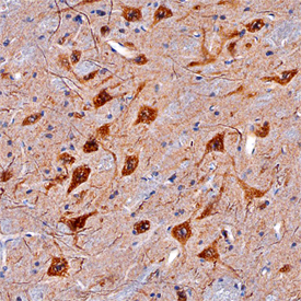
CDNF in Mouse Brain. CDNF was detected in perfusion fixed frozen sections of mouse brain (medulla) using Goat Anti-Mouse CDNF Antigen Affinity-purified Polyclonal Antibody (Catalog # AF5187) at 5 µg/mL overnight at 4 °C. Tissue was stained using the Anti-Goat HRP-DAB Cell & Tissue Staining Kit (brown; Catalog # CTS008) and counterstained with hematoxylin (blue). Specific staining was localized to neuronal cell bodies and processes. View our protocol for Chromogenic IHC Staining of Frozen Tissue Sections.
Reconstitution Calculator
Preparation and Storage
- 12 months from date of receipt, -20 to -70 degreesC as supplied. 1 month, 2 to 8 degreesC under sterile conditions after reconstitution. 6 months, -20 to -70 degreesC under sterile conditions after reconstitution.
Background: CDNF
CDNF (conserved dopamine neurotrophic factor), also called Armetl1 (arginine-rich, mutated in early stage tumors-like 1), is a 17-19 kDa secreted protein that shares 52% amino acid (aa) identity with mouse MANF (mesencephalic-astrocyte-derived neurotrophic factor), also called Armet (1). The Armet designation is not preferred, because the proteins as translated are not actually arginine-rich (1). However, both CDNF and MANF have a high proportion of charged residues, a pattern of eight cysteines shown to form intramoleculular disulfide bonds, and a C-terminal endoplasmic reticulum retention signal (shown to function in MANF) (1-3). The mouse CDNF cDNA encodes a 187 aa protein with a 24 aa signal sequence and a 163 mature sequence (1). Mature mouse CDNF shares 80%, 87%, 83%, and 82% aa identity with human, rat, equine and bovine CDNF, respectively. Although CDNF mRNA and protein are expressed in pre and postnatal mouse brain, they are mostly abundant in adult heart, skeletal muscle and testis. Transcripts within the postnatal mouse brain are concentrated in the hippocampus, thalamus, corpus callosum and optic nerve (1). Like MANF and GDNF, CDNF promotes survival of dopaminergic neurons in vitro (1, 4). In a rat Parkinson's disease model, CDNF also promotes rescue and restoration of dopaminergic neurons in vivo (1).
- Lindholm, P. et al. (2007) Nature 448:73.
- Mizobuchi, N. et al. (2007) Cell Struct. Funct. 32:41.
- Raykhel, I. et al. (2007) J. Cell Biol. 179:1193.
- Petrova, P. et al. (2003) J. Mol. Neurosci. 20:173.
Product Datasheets
Citation for Mouse CDNF Antibody
R&D Systems personnel manually curate a database that contains references using R&D Systems products. The data collected includes not only links to publications in PubMed, but also provides information about sample types, species, and experimental conditions.
1 Citation: Showing 1 - 1
-
CDNF Interacts with ER Chaperones and Requires UPR Sensors to Promote Neuronal Survival
Authors: A Eesmaa, LY Yu, H Göös, T Danilova, K Nõges, E Pakarinen, M Varjosalo, M Lindahl, P Lindholm, M Saarma
International Journal of Molecular Sciences, 2022-08-22;23(16):.
Species: Mouse
Sample Types: Tissue Homogenates
Applications: ELISA Capture
FAQs
No product specific FAQs exist for this product, however you may
View all Antibody FAQsReviews for Mouse CDNF Antibody
There are currently no reviews for this product. Be the first to review Mouse CDNF Antibody and earn rewards!
Have you used Mouse CDNF Antibody?
Submit a review and receive an Amazon gift card.
$25/€18/£15/$25CAN/¥75 Yuan/¥2500 Yen for a review with an image
$10/€7/£6/$10 CAD/¥70 Yuan/¥1110 Yen for a review without an image

