Mouse CXCR7/RDC-1 Antibody Summary
Applications
Please Note: Optimal dilutions should be determined by each laboratory for each application. General Protocols are available in the Technical Information section on our website.
Scientific Data
 View Larger
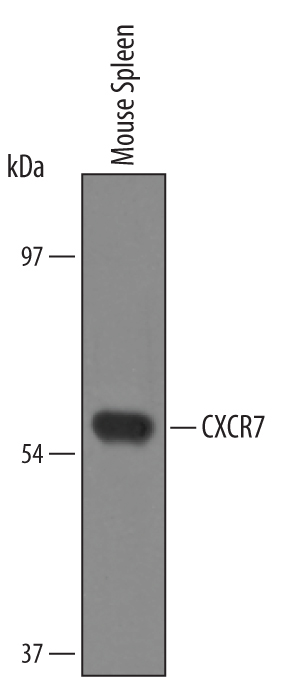
Detection of Mouse CXCR7/RDC‑1 by Western Blot. Western blot shows lysates of mouse spleen tissue. PVDF Membrane was probed with 1 µg/mL of Mouse CXCR7/RDC-1 Antigen Affinity-purified Polyclonal Antibody (Catalog # AF4227) followed by HRP-conjugated Anti-Sheep IgG Secondary Antibody (Catalog # HAF016). A specific band was detected for CXCR7/RDC-1 at approximately 60 kDa (as indicated). This experiment was conducted under reducing conditions and using Immunoblot Buffer Group 8.
 View Larger
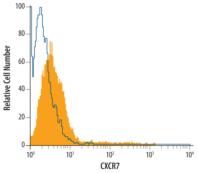
Detection of CXCR7/RDC‑1 in Mouse Neural Progenitor Cells by Flow Cytometry. Undifferentiated mouse neural progenitor cells were stained with Sheep Anti-Mouse CXCR7/RDC-1 Antigen Affinity-purified Polyclonal Antibody (Catalog # AF4227, filled histogram) or control antibody (Catalog # 5-001-A, open histogram), followed by NorthernLights™ 557-conjugated Anti-Sheep IgG Secondary Antibody (Catalog # NL010).
Reconstitution Calculator
Preparation and Storage
- 12 months from date of receipt, -20 to -70 degreesC as supplied. 1 month, 2 to 8 degreesC under sterile conditions after reconstitution. 6 months, -20 to -70 degreesC under sterile conditions after reconstitution.
Background: CXCR7/RDC-1
CXCR7 (CXC chemokine receptor 7; also GPRN1, RDC1 and chemokine orphan receptor 1) is a 60 kDa member of the G-protein coupled receptor 1 family. It is expressed on multiple cell types, including neurons, T cells, NK cells, neutrophils, B cells plus angiogenic endothelial cells. CXCR7 forms both homodimers and heterodimers with CXCR4. It selectively binds I-TAC and SDF1, and appears to involve beta -arrestin2 during signaling. Notably, a CXCR7:CXCR4 heterodimer shows increased responsiveness to SDF1, and I-TAC may actually block some SDF1-mediated migration activity. Mouse CXCR7 is a 7-transmembrane glycoprotein that is 362 amino acids (aa) in length. It contains a 47 aa N-terminal extracellular region plus a 43 aa C-terminal cytoplasmic domain. Over aa 1-47, 103-118, 184-213 and 274-296 collectively, mouse CXCR7 shares 97% and 91% aa identity with rat and human CXCR7, respectively.
Product Datasheets
Citations for Mouse CXCR7/RDC-1 Antibody
R&D Systems personnel manually curate a database that contains references using R&D Systems products. The data collected includes not only links to publications in PubMed, but also provides information about sample types, species, and experimental conditions.
2
Citations: Showing 1 - 2
Filter your results:
Filter by:
-
The role of SDF-1-CXCR4/CXCR7 axis in the therapeutic effects of hypoxia-preconditioned mesenchymal stem cells for renal ischemia/reperfusion injury.
Authors: Liu H, Liu S, Li Y, Wang X, Xue W, Ge G, Luo X
PLoS ONE, 2012-04-12;7(4):e34608.
Species: Mouse
Sample Types: Whole Cells
Applications: Neutralization -
Hypoxic preconditioning advances CXCR4 and CXCR7 expression by activating HIF-1alpha in MSCs.
Authors: Liu H, Xue W, Ge G, Luo X, Li Y, Xiang H, Ding X, Tian P, Tian X
Biochem. Biophys. Res. Commun., 2010-09-24;401(4):509-15.
Species: Mouse
Sample Types: Cell Lysates, Whole Cells
Applications: Neutralization, Western Blot
FAQs
No product specific FAQs exist for this product, however you may
View all Antibody FAQsReviews for Mouse CXCR7/RDC-1 Antibody
There are currently no reviews for this product. Be the first to review Mouse CXCR7/RDC-1 Antibody and earn rewards!
Have you used Mouse CXCR7/RDC-1 Antibody?
Submit a review and receive an Amazon gift card.
$25/€18/£15/$25CAN/¥75 Yuan/¥2500 Yen for a review with an image
$10/€7/£6/$10 CAD/¥70 Yuan/¥1110 Yen for a review without an image


