Mouse
EpCAM/TROP-1 Antibody Summary
Accession # Q99JW5
Applications
Please Note: Optimal dilutions should be determined by each laboratory for each application. General Protocols are available in the Technical Information section on our website.
Scientific Data
 View Larger
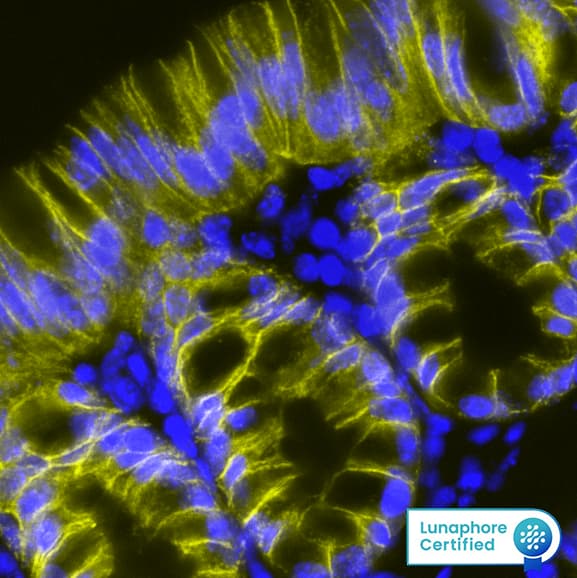
Detection of EPCAM in Mouse Colon via seqIF™ staining on COMET™ EPCAM Antibody was detected in immersion fixed paraffin-embedded sections of mouse Colon using Rat Anti-Mouse EPCAM, Monoclonal Antibody (Catalog # MAB11623) at 20ug/mL at 37 ° Celsius for 4 minutes. Before incubation with the primary antibody, tissue underwent an all-in-one dewaxing and antigen retrieval preprocessing using PreTreatment Module (PT Module) and Dewax and HIER Buffer H (pH 9; Epredia Catalog # TA-999-DHBH). Tissue was stained using the Alexa Fluor™ 647 Goat anti-Rat IgG Secondary Antibody at 1:200 at 37°Celsius for 2 minutes. (Yellow; Lunaphore Catalog # DR647RT) and counterstained with DAPI (blue; Lunaphore Catalog # DR100). Specific staining was localized to the membrane. Protocol available in COMET™ Panel Builder.
 View Larger
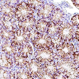
Detection of EpCAM/TROP‑1 in Mouse Colon. EpCAM/TROP‑1 was detected in immersion fixed paraffin-embedded sections of mouse colon using Rat Anti-Mouse EpCAM/TROP‑1 Monoclonal Antibody (Catalog # MAB11623) at 5 µg/ml overnight at 4 °C. Before incubation with the primary antibody, tissue was subjected to heat-induced epitope retrieval using VisUCyte Antigen Retrieval Reagent-Basic (Catalog # VCTS021). Tissue was stained using the HRP-conjugated Anti-Rat IgG Secondary Antibody (Catalog # HAF005) and counterstained with hematoxylin (blue). Specific staining was localized to the membrane of glandular epithelial cells. View our protocol for Chromogenic IHC Staining of Paraffin-embedded Tissue Sections.
 View Larger
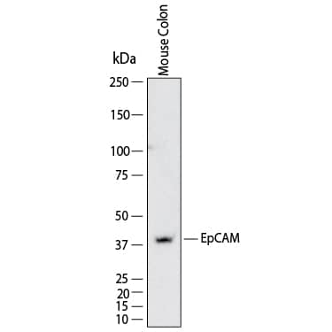
Detection of Mouse EpCAM/TROP‑1 by Western Blot. Western Blot shows lysates of mouse colon tissue. PVDF membrane was probed with 4 µg/ml of Rat Anti-Mouse EpCAM/TROP‑1 Monoclonal Antibody (Catalog # MAB11623) followed by HRP-conjugated Anti-Rat IgG Secondary Antibody (Catalog # HAF005). A specific band was detected for EpCAM/TROP‑1 at approximately 40 kDa (as indicated). This experiment was conducted under reducing conditions and using Western Blot Buffer Group 1.
Reconstitution Calculator
Preparation and Storage
- 12 months from date of receipt, -20 to -70 °C as supplied.
- 1 month, 2 to 8 °C under sterile conditions after reconstitution.
- 6 months, -20 to -70 °C under sterile conditions after reconstitution.
Background: EpCAM/TROP1
Epithelial Cellular Adhesion Molecule (EpCAM), also known as EGP314 (Epithelial glycoprotein 314), TACSTD1 (tumor-associated calcium signal transducer 1) and CD326 is a 292 amino acid (aa), 40 kDa transmembrane glycoprotein composed of a 243 aa extracellular domain with two epidermal-growth-factor-like (EGF-like) repeats within the cysteine-rich N-terminal region, a 23 aa transmembrane domain, and a 26 aa cytoplasmic domain. Human and mouse EpCAM share 82% aa sequence identity. During embryonic development, EpCAM is detected in fetal lung, kidney, liver, pancreas, skin, and germ cells. EpCAM has been shown function as a homophilic Ca2+ independent adhesion molecule (1). Homophilic adhesion via EpCAM requires the interaction of both EGF-like repeats, with the first EGF-like repeat mediating reciprocal interaction between EpCAM molecules on opposing cells, while the second repeat is involved in lateral interaction of EpCAM. Lateral interaction of EpCAM lead to the formation of dimers and tetramers (2). During homophilic adhesion the cytoplasmic tail of EpCAM interacts with the actin cytoskeleton via a direct association alpha -actinin (3).
- Litvinow, S.V. et al. (1994) J. Cell Biol. 125:437.
- Balzar, M. et al. (2001) Mol. Cell. Biol. 21:2570.
- Balzar, M. et al. (1998) Mol. Cell. Biol. 18:4388.
Product Datasheets
FAQs
No product specific FAQs exist for this product, however you may
View all Antibody FAQsReviews for Mouse
EpCAM/TROP-1 Antibody
There are currently no reviews for this product. Be the first to
review Mouse
EpCAM/TROP-1 Antibody and earn rewards!
Have you used Mouse
EpCAM/TROP-1 Antibody?
Submit a review and receive an Amazon gift card.
$25/€18/£15/$25CAN/¥75 Yuan/¥2500 Yen for a review with an image
$10/€7/£6/$10 CAD/¥70 Yuan/¥1110 Yen for a review without an image


