Mouse G-CSF Antibody Summary
Val31-Ala208
Accession # P09920
Applications
This antibody functions as an ELISA detection antibody when paired with Rat Anti-Mouse G‑CSF Monoclonal Antibody (Catalog # MAB414R).
This product is intended for assay development on various assay platforms requiring antibody pairs. We recommend the Mouse G-CSF DuoSet ELISA Kit (Catalog # DY414) for convenient development of a sandwich ELISA or the Mouse G-CSF Quantikine ELISA Kit (Catalog # MCS00) for a complete optimized ELISA.
Please Note: Optimal dilutions should be determined by each laboratory for each application. General Protocols are available in the Technical Information section on our website.
Scientific Data
 View Larger
Cell Proliferation Induced by G‑CSF and Neutralization by Mouse G‑CSF Antibody. Recombinant Mouse G-CSF (Catalog # 414-CS) stimulates proliferation in the NFS-60 mouse myeloid cell line in a dose-dependent manner (orange line). Proliferation elicited by Recombinant Mouse G-CSF (0.125 ng/mL) is neutralized (green line) by increasing concentrations of Goat Anti-Mouse G-CSF Antigen Affinity-purified Polyclonal Antibody (Catalog # AF-414-NA). The ND50 is typically 0.0025-0.0075 µg/mL.
 View Larger
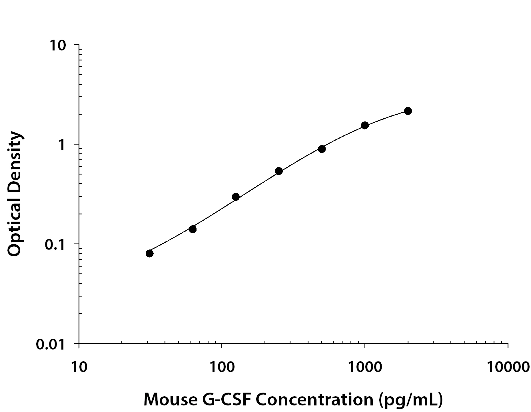
Mouse G‑CSF ELISA Standard Curve. Recombinant Mouse G-CSF protein was serially diluted 2-fold and captured by Rat Anti-Mouse G-CSF Monoclonal Antibody (Catalog # MAB414R) coated on a Clear Polystyrene Microplate (Catalog # DY990). Goat Anti-Mouse G-CSF Antigen Affinity-purified Polyclonal Antibody (Catalog # AF-414-NA) was biotinylated and incubated with the protein captured on the plate. Detection of the standard curve was achieved by incubating Streptavidin-HRP (Catalog # DY998) followed by Substrate Solution (Catalog # DY999) and stopping the enzymatic reaction with Stop Solution (Catalog # DY994).
Reconstitution Calculator
Preparation and Storage
- 12 months from date of receipt, -20 to -70 °C as supplied.
- 1 month, 2 to 8 °C under sterile conditions after reconstitution.
- 6 months, -20 to -70 °C under sterile conditions after reconstitution.
Background: G-CSF
G-CSF is a pleiotropic cytokine best known for its specific effects on the proliferation, differentiation, and activation of hematopoietic cells of the neutrophilic granulocyte lineage. It is produced mainly by monocytes and macrophages upon activation by endotoxin, TNF-alpha and IFN-gamma. Other cell types including fibroblasts, endothelial cells, astrocytes and bone marrow stromal cells can also secrete G-CSF after LPS, IL-1 or TNF-alpha activation. In addition, various carcinoma cell lines and myeloblastic leukemia cells can express G-CSF constitutively.
The murine G-CSF cDNA encodes a 208 amino acid (aa) residue precursor protein containing a 30 aa residue signal peptide that is proteolytically cleaved to generate the 178 aa residue mature protein. Human G-CSF is 73% identical at the amino acid level to murine G-CSF and the two proteins show species cross-reactivity.
In vitro, G-CSF stimulates growth, differentiation and functions of cells from the neutrophil lineage. It also has blast cell growth factor activity and can synergize with IL-3 to shorten the Go period of early hematopoietic progenitors. Consistent with its in vitro functions, G-CSF has been found to play important roles in defense against infection, in inflammation and repair, and in the maintenance of steady state hematopoiesis.
Product Datasheets
Citations for Mouse G-CSF Antibody
R&D Systems personnel manually curate a database that contains references using R&D Systems products. The data collected includes not only links to publications in PubMed, but also provides information about sample types, species, and experimental conditions.
2
Citations: Showing 1 - 2
Filter your results:
Filter by:
-
G-CSF promotes the proliferation of developing cardiomyocytes in vivo and in derivation from ESCs and iPSCs.
Authors: Shimoji K, Yuasa S, Onizuka T, Hattori F, Tanaka T, Hara M, Ohno Y, Chen H, Egasgira T, Seki T, Yae K, Koshimizu U, Ogawa S, Fukuda K
Cell Stem Cell, 2010-03-05;6(3):227-37.
Species: Mouse
Sample Types: Whole Cells
Applications: Neutralization -
Bv8 regulates myeloid-cell-dependent tumour angiogenesis.
Authors: Shojaei F, Wu X, Zhong C, Yu L, Liang XH, Yao J, Blanchard D, Bais C, Peale FV, van Bruggen N, Ho C, Ross J, Tan M, Carano RA, Meng YG, Ferrara N
Nature, 2007-12-06;450(7171):825-31.
Species: Mouse
Sample Types: Cell Lysates
Applications: Neutralization
FAQs
No product specific FAQs exist for this product, however you may
View all Antibody FAQsReviews for Mouse G-CSF Antibody
Average Rating: 1 (Based on 1 Review)
Have you used Mouse G-CSF Antibody?
Submit a review and receive an Amazon gift card.
$25/€18/£15/$25CAN/¥75 Yuan/¥2500 Yen for a review with an image
$10/€7/£6/$10 CAD/¥70 Yuan/¥1110 Yen for a review without an image
Filter by:
Saw no bands on western blot.


