Mouse Galectin-1 Antibody Summary
Ala2-Glu135
Accession # P16045
Applications
Please Note: Optimal dilutions should be determined by each laboratory for each application. General Protocols are available in the Technical Information section on our website.
Scientific Data
 View Larger
Detection of Mouse Galectin‑1 by Simple WesternTM. Simple Western lane view shows lysates of C2C12 mouse myoblast cell line, loaded at 0.2 mg/mL. A specific band was detected for Galectin‑1 at approximately 18 kDa (as indicated) using 50 µg/mL of Rat Anti-Mouse Galectin‑1 Monoclonal Antibody (Catalog # MAB1245) followed by 1:50 dilution of HRP-conjugated Anti-Rat IgG Secondary Antibody (Catalog # HAF005). This experiment was conducted under reducing conditions and using the 12-230kDa separation system.
 View Larger
Detection of Mouse Galectin‑1 by Western Blot. Western blot shows lysates of C2C12 mouse myoblast cell line. PVDF membrane was probed with 2 µg/mL of Rat Anti-Mouse Galectin‑1 Monoclonal Antibody (Catalog # MAB1245) followed by HRP-conjugated Anti-Rat IgG Secondary Antibody (Catalog # HAF005). A specific band was detected for Galectin‑1 at approximately 14 kDa (as indicated). This experiment was conducted under reducing conditions and using Western Blot Buffer Group 1.
Reconstitution Calculator
Preparation and Storage
- 12 months from date of receipt, -20 to -70 °C as supplied.
- 1 month, 2 to 8 °C under sterile conditions after reconstitution.
- 6 months, -20 to -70 °C under sterile conditions after reconstitution.
Background: Galectin-1
The galectins constitute a large family of carbohydrate-binding proteins with specificity for N-acetyl-lactosamine-containing glycoproteins. At least 14 mammalian galectins, which share structural similarities in their carbohydrate recognition domains (CRD), have been identified to date. The galectins have been classified into the prototype galectins (-1, -2, -5, -7, -10, -11, -13, -14), which contain one CRD and exist either as a monomer or a noncovalent homodimer; the chimera galectins (Galectin-3) containing one CRD linked to a nonlectin domain; and the tandem-repeat galectins (-4, -6, -8, -9, -12) consisting of two CRDs joined by a linker peptide. Galectins lack a classical signal peptide and can be localized to the cytosolic compartments where they have intracellular functions. However, via one or more as yet unidentified non-classical secretory pathways, galectins can also be secreted to function extracellularly. Individual members of the galectin family have different tissue distribution profiles and exhibit subtle differences in their carbohydrate-binding specificities. Each family member may preferentially bind to a unique subset of cell-surface glycoproteins (1-4). Mouse Galectin-1, also known as beta-galactoside-binding lectin L-14-I, lactose-binding lectin 1, S-Lac lectin 1, galaptin and 14 kDa lectin, is a monomeric or homodimeric prototype galectin that is expressed in a variety of cells and tissues including muscle, heart, lymph nodes, spleen, thymus, macrophages, B cells, T cells, dendritic cells, and tumor cells. It preferentially binds laminin, fibronectin, 90K/Mac-2BP, CD45, CD43, CD7, CD2, CD3, and ganglioside GM1. Galectin-1 modulates cell growth, proliferation and differentiation, either positively or negatively, depending on the cell type and activation status. It controls cell survival by inducing apoptosis of activated T cells and immature thymocytes. It modulates cytokine secretion by inducing Th2 type cytokines and inhibiting pro-inflammatory cytokine production. Galectin-1 can also modulate cell-cell as well as cell-matrix interactions and depending on the cell type and developmental stage, promote cell attachment or detachment. Galectin-1 has immunosuppressive and anti-inflammatory properties and has been shown to suppress acute and chronic inflammation and autoimmunity. Mouse and human Galectin-1 share about 88% amino acid sequence similarity (1-5).
- Rabinovich, A. et al. (2002) Trends in Immunol. 23:313.
- Rabinovich, A. et al. (2002) J. Leukocyte Biology 71:741.
- Hughes, R.C. (2001) Biochimie 83:667.
- R&D Systems’ Cytokine Bulletin, Summer (2002).
- Goldring, K. et al. (2002) J. Cell Science 115:355.
Product Datasheets
Citation for Mouse Galectin-1 Antibody
R&D Systems personnel manually curate a database that contains references using R&D Systems products. The data collected includes not only links to publications in PubMed, but also provides information about sample types, species, and experimental conditions.
1 Citation: Showing 1 - 1
-
Identification of a lectin causing the degeneration of neuronal processes using engineered embryonic stem cells.
Authors: Plachta N, Annaheim C, Bissiere S, Lin S, Ruegg M, Hoving S, Muller D, Poirier F, Bibel M, Barde YA
Nat. Neurosci., 2007-05-07;10(6):712-9.
Species: Mouse
Sample Types: Cell Lysates
Applications: Western Blot
FAQs
No product specific FAQs exist for this product, however you may
View all Antibody FAQsReviews for Mouse Galectin-1 Antibody
Average Rating: 4.7 (Based on 3 Reviews)
Have you used Mouse Galectin-1 Antibody?
Submit a review and receive an Amazon gift card.
$25/€18/£15/$25CAN/¥75 Yuan/¥1250 Yen for a review with an image
$10/€7/£6/$10 CAD/¥70 Yuan/¥1110 Yen for a review without an image
Filter by:
Works at 1:1000
Specificity: Specific
Sensitivity: Sensitive
Buffer: BSA
Dilution: 1:1000
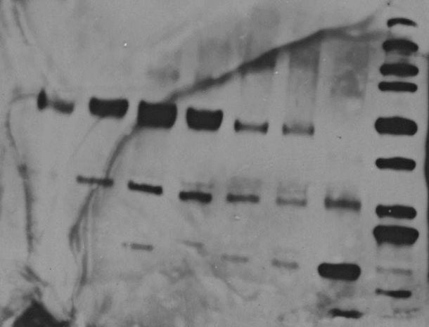
This antibody is very sensitive to detect galectin-1, last lane is marker, last but one is purified galectin, and the rest lanes are cell lysates from differnet cancer cell lines, so it shows that galectin-1 is actually modified.
Only the last bands at bottom is galectin-1, the rest bands are IgG.
Specificity: Specific
Sensitivity: Sensitive
Buffer: BSA
Dilution: 1:1000


