Mouse L1CAM PE-conjugated Antibody Summary
Applications
Please Note: Optimal dilutions should be determined by each laboratory for each application. General Protocols are available in the Technical Information section on our website.
Scientific Data
 View Larger
Detection of L1CAM in Mouse Splenocytes by Flow Cytometry. Mouse splenocytes were stained with Rat Anti-Mouse L1CAM PE-conjugated Monoclonal Antibody (Catalog # FAB5674P, filled histogram) or isotype control antibody (IC006P, open histogram). View our protocol for Staining Membrane-associated Proteins.
 View Larger
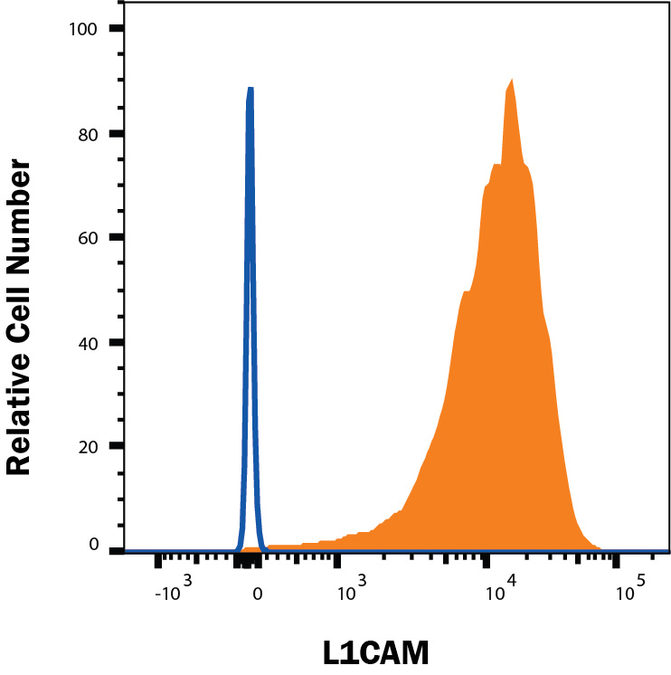
Detection of L1CAM in Neuro-2A cells by Flow Cytometry. Neuro-2A cells were stained with Rat Anti-Mouse L1CAM PE‑conjugated Monoclonal Antibody (Catalog # FAB5674P, filled histogram) or isotype control antibody (Catalog # IC006P, open histogram). View our protocol for Staining Membrane-associated Proteins.
Reconstitution Calculator
Preparation and Storage
Background: L1CAM
L1CAM, also known as Neural Cell Adhesion Molecule L1 (NCAM-L1) and CD171, is a 200‑220 kDa type I transmembrane glycoprotein belonging to the immunoglobulin superfamily, L1/Neurofascin/NgCAM family of molecules. L1CAM is expressed by neurons, especially by growing axons on their growth cones. Non-neuronal cells such as Schwann cells, astrocytes, epithelial cells, and cells of myelomonocytic and lymphoid origin also express L1CAM. Mature mouse L1CAM consists of a 1104 amino acid (aa) extracellular domain (ECD) with 6 Ig-like domains and 5 fibronectin type-III domains, a 23 aa transmembrane region, and a 114 aa cytoplasmic tail. Mouse L1CAM shares 88% aa sequence identity with human L1CAM. L1CAM is critical for neural development. Specifically, L1CAM plays a key role in neuronal cell migration, axon outgrowth, axon fasciculation, synaptogenesis, and myelination. L1CAM mediates homophilic cell-cell interaction but also binds heterophilically with Axonin-1, CD24, CD9, Neurocan and several Integrins. L1CAM can undergo membrane‑proximal cleavage by ADAM10 and ADAM17, leading to the release of the soluble ECD and the generation of a membrane‑bound stub. The soluble ECD can serve as a substrate for integrin-mediated cell adhesion, thereby stimulating cellular motility and cell migration. L1CAM also plays a role in the ontogeny of human tumors, and its expression is linked to poor prognosis. Proteolytic processing by ADAM10 and presenilin/ gamma -secretase is essential for "forward signaling" where an L1CAM intracellular domain translocates to the nucleus and participates in gene regulation. Defects in L1CAM are the cause of the neurological MASA/CRASH syndrome. In addition, uncleaved L1CAM can cluster with Integrin alpha 5 either in-cis, or in-trans, inducing IL-1 beta expression and subsequent NF- kappa B activation. This is referred to as "reverse signaling".
Product Datasheets
FAQs
No product specific FAQs exist for this product, however you may
View all Antibody FAQsReviews for Mouse L1CAM PE-conjugated Antibody
Average Rating: 4 (Based on 1 Review)
Have you used Mouse L1CAM PE-conjugated Antibody?
Submit a review and receive an Amazon gift card.
$25/€18/£15/$25CAN/¥75 Yuan/¥2500 Yen for a review with an image
$10/€7/£6/$10 CAD/¥70 Yuan/¥1110 Yen for a review without an image
Filter by:


