Mouse MMR/CD206 PE-conjugated Antibody Summary
Leu19-Ala1388
Accession # Q2HZ94
Applications
Please Note: Optimal dilutions should be determined by each laboratory for each application. General Protocols are available in the Technical Information section on our website.
Scientific Data
 View Larger
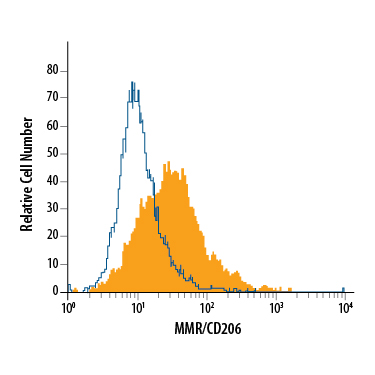
Detection of MMR/CD206 in J774A.1 Mouse Cell Line by Flow Cytometry. J774A.1 mouse reticulum cell sarcoma macrophage cell line was stained with Goat Anti-Mouse MMR/CD206 PE-conjugated Antigen Affinity-purified Polyclonal Antibody (Catalog # FAB2535P, filled histogram) or isotype control antibody (Catalog # IC108P, open histogram). View our protocol for Staining Membrane-associated Proteins.
Reconstitution Calculator
Preparation and Storage
- 12 months from date of receipt, 2 to 8 °C as supplied.
Background: MMR/CD206
The mouse Macrophage Mannose Receptor (MMR), also known as CD206 and MRC1 (mannose receptor C, type 1), is a 175 kDa scavenger receptor that is expressed on tissue macrophages, myeloid dendritic cells, and liver and lymphatic endothelial cells (1). It belongs to a family of receptors sharing similar protein structure that also includes DEC205, phospholipase A2 receptor, and Endo180 (2, 3). The mouse MMR protein is synthesized as a 1456 amino acid (aa) precursor that contains a 19 aa signal sequence, a 1369 aa extracellular region, a 21 aa transmembrane segment and a 47 aa cytoplasmic domain (4). Its extracellular region is composed of an N-terminal cysteine-rich domain, followed by a single fibronectin type II repeat, and eight C-type lectin carbohydrate recognition domains (CRD) (3‑5). Mouse to human, the extracellular region is 82% aa identical. The cysteine-rich domain mediates recognition of sulfated N-acetylgalactosamine, which occurs on some extracellular matrix proteins and is the terminal sugar of the unusual oligosaccharides present on pituitary hormones such as lutropin and thyrotropin (6). Several of the CRDs participate in the Ca2+-dependent recognition of carbohydrates showing a preference for branched sugars with terminal mannose, fucose or N‑acetylglucosamine (7). The cytoplasmic domain of MMR includes a tyrosine-based motif for internalization in clathrin-coated vesicles. Once internalized, ligands are released following acidification of phagosomes or endosomes, and the receptor recycles to the cell surface (3, 8). MMR mediates phagocytosis upon binding to target structures that occur on a variety of pathogenic microorganisms including Gram-negative and Gram-positive bacteria, yeasts, parasites, and mycobacteria. MMR also functions to maintain homeostasis through the endocytosis of potentially harmful glycoproteins associated with inflammation (2, 3).
- East, L. and C. Isake (2002) Biochim. Biophys. Acta 1572:364.
- Chieppa, M. et al. (2003) J. Immunol. 171:4552.
- Figdor, C. et al. (2002) Nat. Rev. Immunol. 2:77.
- Harris, N. et al. (1992) Blood 80:2363.
- Taylor, M. et al. (1990) J. Biol. Chem. 265:12156.
- Leteux, C. et al. (2000) J. Exp. Med. 191:1117.
- Martinez-Pomares, L. et al. (2001) Immunobiology 204:527.
- Feinberg, H. et al. (2000) J. Biol. Chem. 275:21539.
Product Datasheets
Citations for Mouse MMR/CD206 PE-conjugated Antibody
R&D Systems personnel manually curate a database that contains references using R&D Systems products. The data collected includes not only links to publications in PubMed, but also provides information about sample types, species, and experimental conditions.
9
Citations: Showing 1 - 9
Filter your results:
Filter by:
-
Inflammasome Coordinates Senescent Chronic Wound Induced by Thalassophryne nattereri Venom
Authors: Lima, C;Andrade-Barros, AI;Carvalho, FF;Falc�o, MAP;Lopes-Ferreira, M;
International journal of molecular sciences
Species: Mouse
Sample Types: Whole Cells
Applications: Flow Cytometry -
Chronic exposure to biomass ambient particulate matter triggers alveolar macrophage polarization and activation in the rat lung
Authors: S Wang, Y Chen, W Hong, B Li, Y Zhou, P Ran
Journal of Cellular and Molecular Medicine, 2022-01-06;0(0):.
Species: Rat
Sample Types: Whole Cells
Applications: Flow Cytometry -
Anti-HER2 induced myeloid cell alterations correspond with increasing vascular maturation in a murine model of HER2+ breast cancer
Authors: MJ Bloom, AM Jarrett, TA Triplett, AK Syed, T Davis, TE Yankeelov, AG Sorace
BMC Cancer, 2020-04-28;20(1):359.
Species: Mouse
Sample Types: Whole Cells
Applications: Flow Cytometry -
Tumoral NOX4 recruits M2 tumor-associated macrophages via ROS/PI3K signaling-dependent various cytokine production to promote NSCLC growth
Authors: J Zhang, H Li, Q Wu, Y Chen, Y Deng, Z Yang, L Zhang, B Liu
Redox Biol, 2019-02-06;22(0):101116.
Species: Mouse
Sample Types: Xenograft
Applications: Flow Cytometry -
Deletion of lactate dehydrogenase-A in myeloid cells triggers antitumor immunity
Authors: P Seth, E Csizmadia, A Hedblom, M Vuerich, H Xie, M Li, MS Longhi, B Wegiel
Cancer Res., 2017-04-26;0(0):.
Species: Mouse
Sample Types: Whole Cells
Applications: Flow Cytometry -
Differential Immunometabolic Phenotype in Th1 and Th2 Dominant Mouse Strains in Response to High-Fat Feeding.
Authors: Jovicic N, Jeftic I, Jovanovic I, Radosavljevic G, Arsenijevic N, Lukic M, Pejnovic N
PLoS ONE, 2015-07-28;10(7):e0134089.
Species: Mouse
Sample Types: Whole Cells
Applications: Flow Cytometry -
Obesity alters adipose tissue macrophage iron content and tissue iron distribution.
Authors: Orr J, Kennedy A, Anderson-Baucum E, Webb C, Fordahl S, Erikson K, Zhang Y, Etzerodt A, Moestrup S, Hasty A
Diabetes, 2013-10-15;63(2):421-32.
Species: Mouse
Sample Types: Whole Cells
Applications: Flow Cytometry -
Alveolar epithelial cells are critical in protection of the respiratory tract by secretion of factors able to modulate the activity of pulmonary macrophages and directly control bacterial growth.
Authors: Chuquimia O, Petursdottir D, Periolo N, Fernandez C
Infect Immun, 2012-11-12;81(1):381-9.
Species: Mouse
Sample Types: Whole Cells
Applications: Flow Cytometry -
Human mesenchymal stem/stromal cells cultured as spheroids are self-activated to produce prostaglandin E2 that directs stimulated macrophages into an anti-inflammatory phenotype.
Authors: Ylostalo J, Bartosh T, Coble K, Prockop D
Stem Cells, 2012-10-01;30(10):2283-96.
Species: Mouse
Sample Types: Whole Cells
Applications: Flow Cytometry
FAQs
No product specific FAQs exist for this product, however you may
View all Antibody FAQsReviews for Mouse MMR/CD206 PE-conjugated Antibody
There are currently no reviews for this product. Be the first to review Mouse MMR/CD206 PE-conjugated Antibody and earn rewards!
Have you used Mouse MMR/CD206 PE-conjugated Antibody?
Submit a review and receive an Amazon gift card.
$25/€18/£15/$25CAN/¥75 Yuan/¥2500 Yen for a review with an image
$10/€7/£6/$10 CAD/¥70 Yuan/¥1110 Yen for a review without an image

