Mouse Oncostatin M/OSM Antibody Summary
Ala24-Arg206
Accession # P53347
Applications
Please Note: Optimal dilutions should be determined by each laboratory for each application. General Protocols are available in the Technical Information section on our website.
Scientific Data
 View Larger
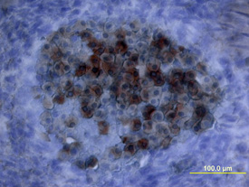
Oncostatin M/OSM in Mouse Embryo. Oncostatin M/OSM was detected in immersion fixed frozen sections of mouse embryo (15 d.p.c, section through spinal cord) using Mouse Oncostatin M/OSM Antigen Affinity-purified Polyclonal Antibody (Catalog # AF-495-NA) at 15 µg/mL overnight at 4 °C. Tissue was stained using the Anti-Goat HRP-DAB Cell & Tissue Staining Kit (brown; Catalog # CTS008) and counterstained with hematoxylin (blue). View our protocol for Chromogenic IHC Staining of Frozen Tissue Sections.
 View Larger
Oncostatin M/OSM in Mouse Embryo. Oncostatin M/OSM was detected in immersion fixed frozen sections of mouse embryo using Mouse Oncostatin M/OSM Antigen Affinity-purified Polyclonal Antibody (Catalog # AF-495-NA) at 15 µg/mL overnight at 4 °C. Tissue was stained using the Anti-Goat HRP-DAB Cell & Tissue Staining Kit (brown; Catalog # CTS008) and counterstained with hematoxylin (blue). View our protocol for Chromogenic IHC Staining of Frozen Tissue Sections.
 View Larger
Cell Proliferation Induced by Oncostatin M/OSM and Neutralization by Mouse Oncostatin M/OSM Antibody. Recombinant Mouse Oncostatin M/OSM (Catalog # 495-MO) stimulates proliferation in the NIH‑3T3 mouse embryonic fibroblast cell line in a dose-dependent manner (orange line). Proliferation elicited by Recombinant Mouse Oncostatin M/OSM (15 ng/mL) is neutralized (green line) by increasing concentrations of Mouse Oncostatin M/OSM Antigen Affinity-purified Polyclonal Antibody (Catalog # AF-495-NA). The ND50 is typically 0.6-3.0 µg/mL.
Reconstitution Calculator
Preparation and Storage
- 12 months from date of receipt, -20 to -70 °C as supplied.
- 1 month, 2 to 8 °C under sterile conditions after reconstitution.
- 6 months, -20 to -70 °C under sterile conditions after reconstitution.
Background: Oncostatin M/OSM
Oncostatin M (OSM) is a member of a cytokine subfamily that includes IL-6, IL-11, LIF, CNTF, and cardiotrophin-1. These cytokines have overlapping biological functions and shared receptor components. Mouse OSM was cloned and identified as an immediate early gene induced in various myeloid and lymphoid cell lines by a subset of cytokines including IL-2, IL-3, GM-CSF, and EPO. The mouse OSM cDNA encodes a 263 amino acid residue precursor protein that shows 48% identity with human OSM. Similar to human OSM, the C-terminal region of mouse OSM contains a highly charged region. Deletion of this C-terminal region appears to be essential for the formation of biologically active mOSM.
The biological activity of human OSM has been shown to be mediated either by the LIF/OSM receptor complex composed of gp130 and LIF R alpha or by a human OSM specific receptor composed of gp130 and OSM R alpha. It remains to be determined if the biological activities of mouse OSM can also be mediated by both receptor complexes in mouse cells.
- Yoshimura, A. et al. (1996) The EMBO Journal 15:1055.
- Ray, P. et al. (1996) Endocrinology 137:1151.
- Rose, T.M. and A.G. Bruce (1994) in Guidebook to Cytokines and Their Receptors, N.A. Nicola, editor, Oxford University Press, New York, p. 127.
Product Datasheets
Citations for Mouse Oncostatin M/OSM Antibody
R&D Systems personnel manually curate a database that contains references using R&D Systems products. The data collected includes not only links to publications in PubMed, but also provides information about sample types, species, and experimental conditions.
11
Citations: Showing 1 - 10
Filter your results:
Filter by:
-
Skin basal cell carcinomas assemble a pro-tumorigenic spatially organized and self-propagating Trem2+ myeloid niche
Authors: Haensel, D;Daniel, B;Gaddam, S;Pan, C;Fabo, T;Bjelajac, J;Jussila, AR;Gonzalez, F;Li, NY;Chen, Y;Hou, J;Patel, T;Aasi, S;Satpathy, AT;Oro, AE;
Nature communications
Species: Mouse
Sample Types: In Vivo
Applications: In Vivo -
Essential roles of the cytokine oncostatin M in crosstalk between muscle fibers and immune cells in skeletal muscle after aerobic exercise
Authors: T Komori, Y Morikawa
The Journal of Biological Chemistry, 2022-11-09;0(0):102686.
Species: Mouse
Sample Types: Tissue Homogenates, Whole Tissue
Applications: IHC, Western Blot -
Concomitant Activation of OSM and LIF Receptor by a Dual-Specific hlOSM Variant Confers Cardioprotection after Myocardial Infarction in Mice
Authors: H Lörchner, JM Adrian-Seg, C Waechter, R Wagner, ME Góes, N Brachmann, K Sreenivasa, A Wietelmann, S Günther, N Doll, T Braun, J Pöling
International Journal of Molecular Sciences, 2021-12-29;23(1):.
Species: Mouse
Sample Types: Cell Lysates
Applications: Western Blot -
Neutrophil extracellular traps (NETs) contribute to pathological changes of ocular graft-vs.-host disease (oGVHD) dry eye: Implications for novel biomarkers and therapeutic strategies
Authors: S An, I Raju, B Surenkhuu, JE Kwon, S Gulati, M Karaman, A Pradeep, S Sinha, C Mun, S Jain
Ocul Surf, 2019-04-06;0(0):.
Species: Mouse
Sample Types: In Vivo
Applications: Neutralization -
Anti-OSM Antibody Inhibits Tubulointerstitial Lesion in a Murine Model of Lupus Nephritis
Authors: Q Liu, Y Du, K Li, W Zhang, X Feng, J Hao, H Li, S Liu
Mediators Inflamm., 2017-05-24;2017(0):3038514.
Species: Mouse
Sample Types: In Vivo
Applications: Neutralization -
Oncostatin M overexpression induces skin inflammation but is not required in the mouse model of imiquimod-induced psoriasis-like inflammation
Eur J Immunol, 2016-05-12;0(0):.
Species: Mouse
Sample Types: Whole Tissue
Applications: IHC-Fr -
Oncostatin m, an inflammatory cytokine produced by macrophages, supports intramembranous bone healing in a mouse model of tibia injury.
Authors: Guihard P, Boutet M, Brounais-Le Royer B, Gamblin A, Amiaud J, Renaud A, Berreur M, Redini F, Heymann D, Layrolle P, Blanchard F
Am J Pathol, 2015-01-02;185(3):765-75.
Species: Mouse
Sample Types: Whole Tissue
Applications: IHC -
Oncostatin M acting via OSMR, augments the actions of IL-1 and TNF in synovial fibroblasts.
Authors: Le Goff B, Singbrant S, Tonkin B, Martin T, Romas E, Sims N, Walsh N
Cytokine, 2014-04-22;68(2):101-9.
Species: Mouse
Sample Types: Whole Tissue
Applications: IHC-P -
Activation of NFAT signaling establishes a tumorigenic microenvironment through cell autonomous and non-cell autonomous mechanisms.
Authors: Tripathi, P, Wang, Y, Coussens, M, Manda, K R, Casey, A M, Lin, C, Poyo, E, Pfeifer, J D, Basappa, N, Bates, C M, Ma, L, Zhang, H, Pan, M, Ding, L, Chen, F
Oncogene, 2013-04-29;33(14):1840-9.
Species: Mouse
Sample Types: Whole Tissue
Applications: IHC -
Long term oncostatin M treatment induces an osteocyte-like differentiation on osteosarcoma and calvaria cells.
Authors: Brounais B, David E, Chipoy C, Trichet V, Ferre V, Charrier C, Duplomb L, Berreur M, Redini F, Heymann D, Blanchard F
Bone, 2009-01-03;44(5):830-9.
Species: Mouse, Rat
Sample Types: Cell Lysates
Applications: Neutralization -
Cardiotrophin-1 in choroid plexus and the cerebrospinal fluid circulatory system.
Authors: Gard AL, Gavin E, Solodushko V, Pennica D
Neuroscience, 2004-01-01;127(1):43-52.
Species: Mouse, Rat
Sample Types: Tissue Homogenates
Applications: Western Blot
FAQs
No product specific FAQs exist for this product, however you may
View all Antibody FAQsReviews for Mouse Oncostatin M/OSM Antibody
There are currently no reviews for this product. Be the first to review Mouse Oncostatin M/OSM Antibody and earn rewards!
Have you used Mouse Oncostatin M/OSM Antibody?
Submit a review and receive an Amazon gift card.
$25/€18/£15/$25CAN/¥75 Yuan/¥2500 Yen for a review with an image
$10/€7/£6/$10 CAD/¥70 Yuan/¥1110 Yen for a review without an image

