Mouse Osteopontin/OPN Antibody
Mouse Osteopontin/OPN Antibody Summary
Leu17-Asn294
Accession # Q547B5
Applications
Please Note: Optimal dilutions should be determined by each laboratory for each application. General Protocols are available in the Technical Information section on our website.
Scientific Data
 View Larger
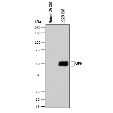
Detection of Mouse Osteopontin/OPN by Western Blot. Western blot shows conditioned media from Neuro-2A mouse neuroblastoma cell line (negative control) and L-929 mouse fibroblast cell line. PVDF membrane was probed with 1 µg/mL of Rabbit Anti-Mouse Osteopontin/OPN Monoclonal Antibody (Catalog # MAB808) followed by HRP-conjugated Anti-Rabbit IgG Secondary Antibody (Catalog # HAF008). A specific band was detected for Osteopontin/OPN at approximately 50 kDa (as indicated). This experiment was conducted under reducing conditions and using Immunoblot Buffer Group 1.
 View Larger
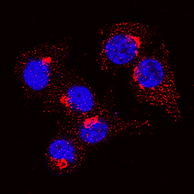
Osteopontin/OPN in RAW 264.7 Mouse Cell Line. Osteopontin/OPN was detected in immersion fixed RAW 264.7 mouse monocyte/macrophage cell line using Rabbit Anti-Mouse Osteopontin/OPN Monoclonal Antibody (Catalog # MAB808) at 3 µg/mL for 3 hours at room temperature. Cells were stained using the NorthernLights™ 557-conjugated Anti-Rabbit IgG Secondary Antibody (red; Catalog # NL004) and counterstained with DAPI (blue). Specific staining was localized to cytoplasm. View our protocol for Fluorescent ICC Staining of Non-adherent Cells.
 View Larger
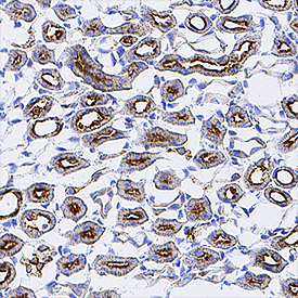
Osteopontin/OPN in Mouse Kidney. Osteopontin/OPN was detected in perfusion fixed frozen sections of mouse kidney using Rabbit Anti-Mouse Osteopontin/OPN Monoclonal Antibody (Catalog # MAB808) at 3 µg/mL for 1 hour at room temperature followed by incubation with the Anti-Rabbit IgG VisUCyte™ HRP Polymer Antibody (Catalog # VC003). Tissue was stained using DAB (brown) and counterstained with hematoxylin (blue). Specific staining was localized to convoluted tubules. View our protocol for IHC Staining with VisUCyte HRP Polymer Detection Reagents.
Reconstitution Calculator
Preparation and Storage
- 12 months from date of receipt, -20 to -70 °C as supplied.
- 1 month, 2 to 8 °C under sterile conditions after reconstitution.
- 6 months, -20 to -70 °C under sterile conditions after reconstitution.
Background: Osteopontin/OPN
Osteopontin (OPN), previously called SPP1 (secreted phosphoprotein 1), Eta-1 (early T lymphocyte activation 1) or BSP (bone sialoprotein), is a secreted molecule in the SIBLING (small integrin-binding ligand N-linked glycoprotein) family of non-collagenous matricellular proteins (1-3). Mouse OPN is synthesized as a 294 amino acid (aa) precursor protein with a 16 aa signal peptide and a 278 aa mature protein (3). Mature mouse OPN shares 79% and 64% aa sequence identity with rat and human OPN, respectively. OPN is highly acidic and has 26 potential Ser/Thr phosphorylation sites and a C‑terminal CD44 binding site (1-4). Depending on tissue-specific modification by O- and N-glycosylation, sulfation, phosphorylation and transglutamination, OPN can be detected at 45-75 kDa (5, 6). The central region of OPN contains RGD and non-RGD binding sites for multiple integrins (3, 4). Adjacent to the RGD motif is the sequence SLAYGLR (SVVYGLR in human) which serves as a cryptic binding site for additional integrins: it is masked in full length OPN but is exposed following OPN cleavage by thrombin in tumors and sites of tissue injury
(6-8). OPN can also be cleaved by MMP-3, -7, -9, and -12 within the SLAYGLR motif and at sites closer to the C-terminus (8, 9). OPN is widely expressed and is prominent in mineralized tissues. It inhibits bone mineralization and kidney stone formation, and promotes inflammation and cell adhesion and migration (1, 2, 4, 6). Its expression is up-regulated during inflammation, obesity, atherosclerosis, cancer, and tissue damage, and contributes to the pathophysiology of these conditions (1, 2, 6, 9, 10).
- Scatena, M. et al. (2007) Arterioscler. Thromb. Vasc. Biol. 27:2302.
- Rangaswami, H. et al. (2006) Trends Cell Biol. 16:79.
- Miyazaki, Y. et al. (1990) J. Biol. Chem. 265:14432.
- Weber, G.F. et al. (2002) J. Leukoc. Biol. 72:752.
- Keykhosravani, M. et al. (2005) Biochemistry 44:6990.
- Kazanecki, C.C. et al. (2007) J. Cell. Biochem. 102:912.
- Senger, D.R. et al. (1994) Mol. Biol. Cell 5:565.
- Yokosaki, Y. et al. (2005) Matrix Biol. 24:418.
- Takafuji, V. et al. (2007) Oncogene 26:6361.
- Kiefer, F.W. et al. (2010) Diabetes 59:935.
Product Datasheets
Citations for Mouse Osteopontin/OPN Antibody
R&D Systems personnel manually curate a database that contains references using R&D Systems products. The data collected includes not only links to publications in PubMed, but also provides information about sample types, species, and experimental conditions.
3
Citations: Showing 1 - 3
Filter your results:
Filter by:
-
Autophagy inhibition prevents lymphatic malformation progression to lymphangiosarcoma by decreasing osteopontin and Stat3 signaling
Authors: F Yang, S Kalantari, B Ruan, S Sun, Z Bian, JL Guan
Nature Communications, 2023-02-22;14(1):978.
Species: Mouse
Sample Types: Cell Lysates
Applications: Western Blot -
Lumenal calcification and microvasculopathy in fetuin-A-deficient mice lead to multiple organ morbidity
Authors: M Herrmann, A Babler, I Moshkova, F Gremse, F Kiessling, U Kusebauch, V Nelea, R Kramann, RL Moritz, MD McKee, W Jahnen-Dec
PLoS ONE, 2020-02-19;15(2):e0228503.
Species: Mouse
Sample Types: Organs, Whole Tissue
Applications: IHC -
Long-term neuronal survival, regeneration, and transient target reconnection after optic nerve crush and mesenchymal stem cell transplantation
Authors: LA Mesentier-, LC Teixeira-P, F Gubert, JF Vasques, AJ Silva-Juni, L Chimeli-Or, G Nascimento, R Mendez-Ote, MF Santiago
Stem Cell Res Ther, 2019-04-17;10(1):121.
Species: Rat
Sample Types: Whole Tissue
Applications: IHC
FAQs
No product specific FAQs exist for this product, however you may
View all Antibody FAQsReviews for Mouse Osteopontin/OPN Antibody
There are currently no reviews for this product. Be the first to review Mouse Osteopontin/OPN Antibody and earn rewards!
Have you used Mouse Osteopontin/OPN Antibody?
Submit a review and receive an Amazon gift card.
$25/€18/£15/$25CAN/¥75 Yuan/¥2500 Yen for a review with an image
$10/€7/£6/$10 CAD/¥70 Yuan/¥1110 Yen for a review without an image


