Mouse/Rat DLL1 Antibody Summary
Gln18-Trp537 (predicted)
Accession # P97677
*Small pack size (-SP) is supplied either lyophilized or as a 0.2 µm filtered solution in PBS.
Applications
Please Note: Optimal dilutions should be determined by each laboratory for each application. General Protocols are available in the Technical Information section on our website.
Scientific Data
 View Larger
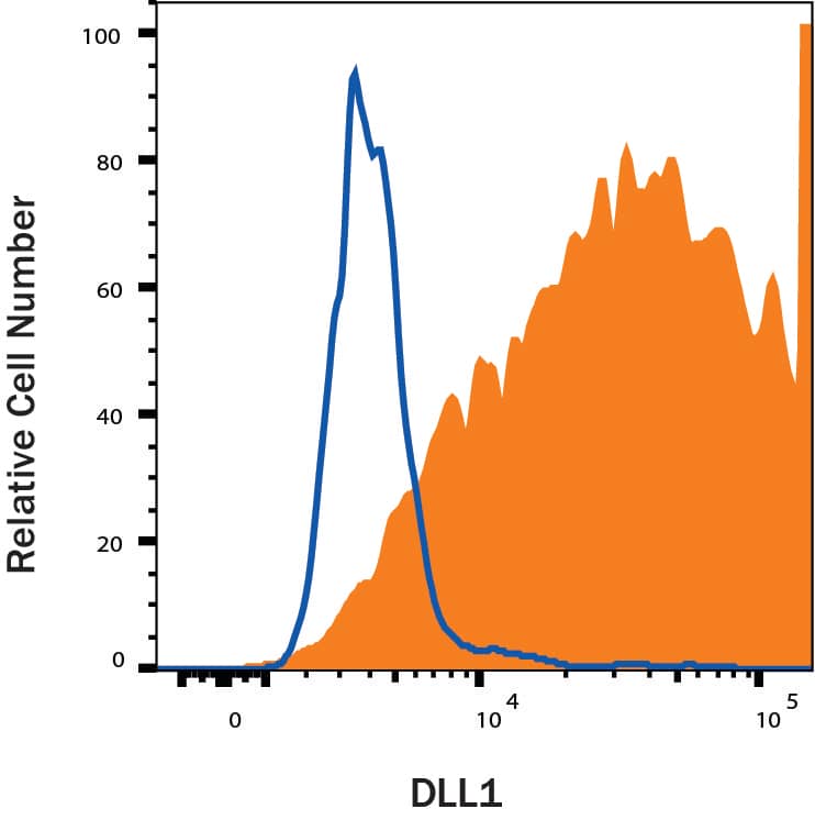
Detection of DLL1 in Mouse Splenocytes by Flow Cytometry. Mouse splenocytes were stained with Sheep Anti-Mouse/Rat DLL1 Antigen Affinity-purified Polyclonal Antibody (Catalog # AF3970, filled histogram) or control antibody (5-001-A, open histogram), followed by NorthernLights™ 637-conjugated Anti-Sheep IgG Secondary Antibody (NL011).
.") View Larger
View Larger
DLL1 in Embryonic Mouse Stomach. DLL1 was detected in immersion fixed frozen sections of embryonic mouse stomach (E13.5) using 10 µg/mL Sheep Anti-Mouse/Rat DLL1 Antigen Affinity-purified Polyclonal Antibody (Catalog # AF3970) overnight at 4 °C. Tissue was stained with the NorthernLights™ 557-conjugated Anti-Sheep IgG Secondary Antibody (red; NL010) and counterstained with DAPI (blue). View our protocol for Fluorescent IHC Staining of Frozen Tissue Sections.
 View Larger
Detection of Mouse DLL1 by Immunocytochemistry/Immunofluorescence Mpdz does not affect cell cell junction assembly.(A, B) HUVECs were either transduced with lentivirus expressing GFP (sh-ctrl) or with lentivirus expressing shRNA against MPDZ (sh-MPDZ). Cells were cultured under sparse conditions (A) or confluent conditions (B). After PFA fixation cells were stained for DLL1 and Nectin-2 or DLL4 and Nectin-2 and counterstained with DAPI. Images were acquired with the confocal microscope LSM 700. Arrow indicates co-localization of DLL1/4 with Nectin-2 at the cell membrane. Arrow head indicates diminished co-localization at the cell membrane. Scale bar: 10 µm. (C) HUVECs were either transfected with control siRNA (si-ctrl) or with siRNA against Nectin-2 (si-Nectin-2). After PFA fixation cells were stained for DLL1 and Nectin-2 or DLL4 and Nectin-2. Images were acquired with the confocal microscope LSM 700. Arrow indicates localization of DLL1/4 at the cell membrane.Scale bar: 10 µm. Image collected and cropped by CiteAb from the following publication (https://pubmed.ncbi.nlm.nih.gov/29620522), licensed under a CC-BY license. Not internally tested by R&D Systems.
 View Larger
Detection of Mouse DLL1 by Western Blot MPDZ promotes Notch signaling activity.(A) HUVECs were either transduced with lentivirus expressing GFP (sh-ctrl) or with lentivirus expressing shRNA against MPDZ (sh-MPDZ). Expression level of Notch target genes HEY1, HEY2 and HES1 were analyzed by qPCR 48 hr after transduction. Data are presented as mean ±SD. n ≥ 3; *, p<0.05; **, p<0.01; ***, p<0.001 unpaired Student’s t-test. (B) Cardiac endothelial cells were isolated from Mpdzfl/fl and Mpdz delta EC mice by magnetic beads bound with CD31 antibodies. Expression levels of Notch target genes Hey1 and Hey2 were analyzed by qPCR. Data are presented as mean ±SD. n = 3; *, p<0.05; ***, p<0.001 unpaired Student’s t-test. (C) HUVECs were either transduced with lentivirus expressing GFP (sh-ctrl) or with lentivirus expressing shRNA against MPDZ (sh-MPDZ). Expression levels of DLL1 and DLL4 were analyzed by immunoblotting 48 hr after transduction. beta -actin served as loading control. Data are presented as mean ±SD. n ≥ 3; n.s., not significant. (D) HUVECs were either transduced with adenovirus expressing GFP (ctrl) or with adenovirus expressing MPDZ. Expression levels of DLL1 and DLL4 were analyzed by immunoblotting 48 hr after transduction. beta -actin served as loading control. Data are presented as mean ±SD. n ≥ 3; n.s., not significant. (E) Lung endothelial cells were isolated from Mpdzfl/fl and Mpdz delta EC mice by CD31 magnetic beads. Protein amounts of Dll1 and Dll4 were analyzed by immunoblotting. beta -actin served as loading control. Data are presented as mean ±SD. n = 3; n.s., not significant.10.7554/eLife.32860.007Figure 2—source data 1.Source data of qantitative PCR analysis related to Figure 2A and B.Source data of qantitative PCR analysis related to Figure 2A and B. Image collected and cropped by CiteAb from the following publication (https://pubmed.ncbi.nlm.nih.gov/29620522), licensed under a CC-BY license. Not internally tested by R&D Systems.
 View Larger
Detection of DLL1 in transfected CHO cells by Flow Cytometry CHO cells transfected with DLL1 were stained with Sheep Anti-Mouse/Rat DLL1 Antigen Affinity-purified Polyclonal Antibody (Catalog # AF3970, filled histogram) or isotype control antibody (Catalog # 5-001-A, open histogram) followed by Allophycocyanin-conjugated Anti-Sheep IgG Secondary Antibody (Catalog # F0127). View our protocol for Staining Membrane-associated Proteins.
Reconstitution Calculator
Preparation and Storage
- 12 months from date of receipt, -20 to -70 °C as supplied.
- 1 month, 2 to 8 °C under sterile conditions after reconstitution.
- 6 months, -20 to -70 °C under sterile conditions after reconstitution.
Background: DLL1
Delta-like protein 1 (DLL1) is a 90-100 kDa type I transmembrane protein in the Delta/Serrate/Lag-2 (DSL) family of Notch ligands. Mature rat DLL1 consists of a 520 amino acid (aa) extracellular domain (ECD) with one DSL domain and eight EGF-like repeats, a 23 aa transmembrane segment, and a 154 aa cytoplasmic domain (1). Within the ECD, rat DLL1 shares 90% and 95% aa sequence identity with human and mouse DLL1, respectively. It shares 26%, 36%, and 53% aa sequence identity with rat DLL2, 3, and 4, respectively. The ADAM9, 12, or 17- mediated proteolysis of DLL1 releases a 60 kDa ECD fragment and regulates the Notch-dependent proliferation of hematopoietic and myogenic progenitor cells (2-4). The residual membrane-bound portion of DLL1 can be cleaved by presenilin-dependent
gamma -secretase, enabling the cytoplasmic domain to migrate to the nucleus (5). DLL1 localizes to adherens junctions on neuronal processes through its association with the scaffolding protein MAGI1 (6). DLL1 is widely expressed, and it plays an important role in embryonic somite formation, cochlear hair cell differentiation, lymphocyte differentiation, and the maintenance of neural and myogenic progenitor cells (4, 7-13). The upregulation of DLL1 in arterial endothelial cells following injury or angiogenic stimulation is central to postnatal arteriogenesis (14). DLL1 is also overexpressed in cervical carcinoma and glioma and contributes to tumor progression (15-16).
- Bettenhausen, B. et al. (1995) Development 121:2407.
- Dyczynska, E. et al. (2007) J. Biol. Chem. 282:436.
- Karanu, F.N. et al. (2001) Blood 97:1960.
- Sun, D. et al. (2008) J. Cell Sci. 121:3815.
- Ikeuchi, T. and S.S. Sisodia (2003) J. Biol. Chem. 278:7751.
- Mizuhara, E. et al. (2005) J. Biol. Chem. 280:26499.
- Takahashi, Y. et al. (2003) Development 130:4259.
- Teppner, I. et al. (2007) BMC Dev. Biol. 7:68.
- Kiernan, A.E. et al. (2005) Development 132:4353.
- Schmitt, T.M. and J.C. Zuniga-Pflucker (2002) Immunity 17:749.
- Hozumi, K. et al. (2004) Nat. Immunol. 5:638.
- Shimojo, H. et al. (2008) Neuron 58:52.
- Schuster-Gossler, K. et al. (2007) Proc. Natl. Acad. Sci. USA 104:537.
- Limbourg, A. et al. (2007) Circ. Res. 100:363.
- Purow, B.W. et al. (2005) Cancer Res. 65:2353.
- Gray, G.E. et al. (1999) Am. J. Pathol. 154:785.
Product Datasheets
Citations for Mouse/Rat DLL1 Antibody
R&D Systems personnel manually curate a database that contains references using R&D Systems products. The data collected includes not only links to publications in PubMed, but also provides information about sample types, species, and experimental conditions.
6
Citations: Showing 1 - 6
Filter your results:
Filter by:
-
Evolution of Cortical Neurogenesis in Amniotes Controlled by Robo Signaling Levels
Authors: Adrián Cárdenas, Ana Villalba, Camino de Juan Romero, Esther Picó, Christina Kyrousi, Athanasia C. Tzika et al.
Cell
-
Lysosomal exocytosis releases pathogenic alpha-synuclein species from neurons in synucleinopathy models
Authors: YX Xie, NN Naseri, J Fels, P Kharel, Y Na, D Lane, J Burré, M Sharma
Nature Communications, 2022-08-22;13(1):4918.
Species: Human
Sample Types: Lysosomes
Applications: ICC -
Multiscale light-sheet organoid imaging framework
Authors: G de Medeiro, R Ortiz, P Strnad, A Boni, F Moos, N Repina, L Challet Me, F Maurer, P Liberali
Nature Communications, 2022-08-18;13(1):4864.
Species: Human
Sample Types: Organoid
Applications: IHC -
MPDZ promotes DLL4-induced Notch signaling during angiogenesis
Authors: F Tetzlaff, MG Adam, A Feldner, I Moll, A Menuchin, J Rodriguez-, D Sprinzak, A Fischer
Elife, 2018-04-05;7(0):.
Species: Mouse
Sample Types: Cell Lysates
Applications: Western Blot -
Cadherin-based adhesions in the apical endfoot are required for active Notch signaling to control neurogenesis in vertebrates.
Authors: Hatakeyama J, Wakamatsu Y, Nagafuchi A, Kageyama R, Shigemoto R, Shimamura K
Development, 2014-04-01;141(8):1671-82.
Species: Chicken, Mouse
Sample Types: Whole Cells, Whole Tissue
Applications: IHC -
Single-cell gene profiling defines differential progenitor subclasses in mammalian neurogenesis.
Authors: Kawaguchi A, Ikawa T, Kasukawa T, Ueda HR, Kurimoto K, Saitou M, Matsuzaki F
Development, 2008-09-01;135(18):3113-24.
Species: Mouse
Sample Types: Whole Tissue
Applications: IHC-Fr
FAQs
No product specific FAQs exist for this product, however you may
View all Antibody FAQsReviews for Mouse/Rat DLL1 Antibody
There are currently no reviews for this product. Be the first to review Mouse/Rat DLL1 Antibody and earn rewards!
Have you used Mouse/Rat DLL1 Antibody?
Submit a review and receive an Amazon gift card.
$25/€18/£15/$25CAN/¥75 Yuan/¥1250 Yen for a review with an image
$10/€7/£6/$10 CAD/¥70 Yuan/¥1110 Yen for a review without an image


