Mouse/Rat Prolactin Antibody Summary
Leu32-Cys228
Accession # NP_035294
*Small pack size (-SP) is supplied either lyophilized or as a 0.2 µm filtered solution in PBS.
Applications
Mouse Prolactin Sandwich Immunoassay
Please Note: Optimal dilutions should be determined by each laboratory for each application. General Protocols are available in the Technical Information section on our website.
Scientific Data
 View Larger
Detection of Mouse and Rat Prolactin by Western Blot. Western blot shows lysates of mouse pituitary tissue and rat pituitary tissue. PVDF membrane was probed with 0.25 µg/mL of Goat Anti-Mouse/Rat Prolactin Antigen Affinity-purified Polyclonal Antibody (Catalog # AF1445) followed by HRP-conjugated Anti-Goat IgG Secondary Antibody (HAF017). A specific band was detected for Prolactin at approximately 23 kDa (as indicated). This experiment was conducted under reducing conditions and using Immunoblot Buffer Group 1.
 View Larger
Cell Proliferation Induced by Prolactin and Neutralization by Mouse Prolactin Antibody. Recombinant Mouse Prolactin (1445-PL) stimulates proliferation in the Nb2-11 rat lymphoma cell line in a dose-dependent manner (orange line). Proliferation elicited by Recombinant Mouse Prolactin (10 ng/mL) is neutralized (green line) by increasing concentrations of Goat Anti-Mouse/Rat Prolactin Antigen Affinity-purified Polyclonal Antibody (Catalog # AF1445). The ND50 is typically 0.25-1.0 µg/mL.
 View Larger
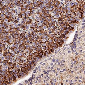
Detection of Prolactin in Mouse Pituitary. Prolactin was detected in immersion fixed paraffin-embedded sections of Mouse Pituitary using Goat Anti-Mouse/Rat Prolactin Antigen Affinity-purified Polyclonal Antibody (Catalog # AF1445) at 1 µg/mL overnight at 4 °C. Tissue was stained using the Anti-Goat IgG VisUCyte™ HRP Polymer Antibody (brown; Catalog # VC004) and counterstained with hematoxylin (blue). Specific staining was localized to cytoplasm in lactotroph cells. View our protocol for IHC Staining with VisUCyte HRP Polymer Detection Reagents.
 View Larger
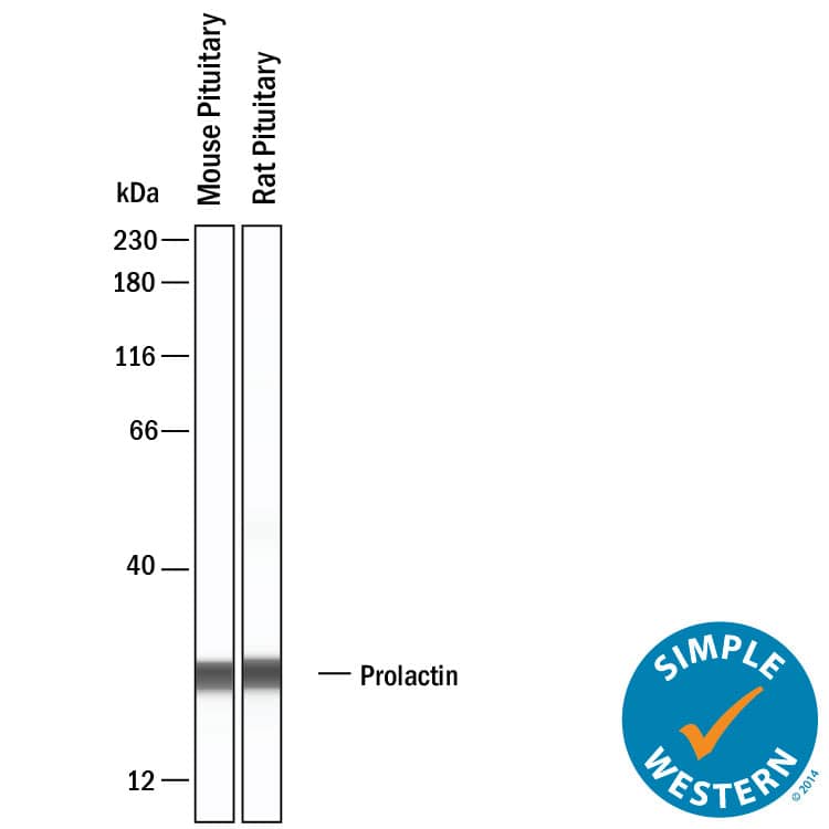
Detection of Mouse and Rat Prolactin by Simple WesternTM. Simple Western shows lysates of mouse pituitary tissue and rat pituitary tissue, loaded at 0.2 mg/ml. A specific band was detected for Prolactin at approximately 26 kDa (as indicated) using 2.5 µg/mL of Goat Anti-Mouse/Rat Prolactin Antigen Affinity-purified Polyclonal Antibody (Catalog # af1445). This experiment was conducted under reducing conditions and using the 12-230kDa separation system.
Reconstitution Calculator
Preparation and Storage
- 12 months from date of receipt, -20 to -70 °C as supplied.
- 1 month, 2 to 8 °C under sterile conditions after reconstitution.
- 6 months, -20 to -70 °C under sterile conditions after reconstitution.
Background: Prolactin
Prolactin (PRL) is a neuroendocrine pituitary hormone. Prolactin is synthesized by the anterior pituitary, placenta, brain, uterus, dermal fibroblasts, decidua, B cells, T cells, NK cells and breast cancer cells. Originally characterized as a lactogenic hormone, further studies have demonstrated broader roles in breast cancer development, regulation of reproductive function, and immunoregulation. In the immune system, Prolactin has been shown to be secreted by human PBMC and to act as a proliferative growth factor. Additionally, Prolactin treatment of human PBMC has been shown to enhance IFN-gamma production. In the breast, Prolactin-induced morphogenesis of the mammary cells is mediated through IGF-2, which in turn upregulates cyclin D1. Prolactin has several molecular forms. The predominant form is a monomer; the non-glycosylated form is 23 kDa and the glycosylated form is 25 kDa. Glycosylated Prolactin is removed from the circulation faster and has been reported to have lower biological potency. Mouse Prolactin cDNA encodes a 228 amino acid (aa) residue protein with a putative 31 aa residue signal peptide. The Prolactin receptor is a transmembrane type I glycoprotein that belongs to the cytokine hematopoietic receptor family. B cells, T cells, macrophages, NK cells, monocytes, CD34+ progenitor cells, neutrophils, mammary gland, liver, kidney, adrenals, ovaries, testis, prostrate, seminal vesicles, and hypothalamus have all been shown to express the Prolactin receptor. Three forms of the receptor, generated by differential splicing, have been identified. These isoforms differ in the length of their cytoplasmic domains. It is believed that the short cytoplasmic form is non-functional. Prolactin signal transduction involves the JAK/STAT families and Src kinase family (1‑9).
- Freeman, M. et al. (2000) Physiological Reviews 80:1523.
- Ben-Johnson, N. et al. (1996) Endoc. Rev. 17:639.
- Cesario, T. et al. (1994) Proc. Soc. Exp. Biol. Med. 205:89.
- Price, A.E. et al. (1995) Endoc. 136:4827.
- Hoffmann, T. et al. (1993) J. Endoc. Invest. 16:807.
- Cole, E. et al. (1991) Endoc. 129:2639.
- Lewis, U. et al. (1985) Endoc. 116:359.
- Matalk, K. (2003) Cytokine 21:187.
- Brisken, C. et al. (2002) Dev. Cell 3:877.
Product Datasheets
Citations for Mouse/Rat Prolactin Antibody
R&D Systems personnel manually curate a database that contains references using R&D Systems products. The data collected includes not only links to publications in PubMed, but also provides information about sample types, species, and experimental conditions.
7
Citations: Showing 1 - 7
Filter your results:
Filter by:
-
MiR-130a-3p Inhibits PRL Expression and Is Associated With Heat Stress-Induced PRL Reduction
Authors: Haojie Zhang, Ting Chen, Jiali Xiong, Baoyu Hu, Junyi Luo, Qianyun Xi et al.
Front Endocrinol (Lausanne)
-
Chemokine CXCL14-like immunoreactivity in the &alphaMSH-producing cells and PRL-producing cells of the flat-tailed house gecko pituitary
Authors: H Suzuki, T Yamamoto
J. Vet. Med. Sci., 2020-02-10;82(4):408-413.
Species: Gecko
Sample Types: Whole Tissue
Applications: IF -
Bone loss caused by dopaminergic degeneration and levodopa treatment in Parkinson's disease model mice
Authors: K Handa, S Kiyohara, T Yamakawa, K Ishikawa, M Hosonuma, N Sakai, A Karakawa, M Chatani, M Tsuji, K Inagaki, Y Kiuchi, M Takami, T Negishi-Ko
Sci Rep, 2019-09-24;9(1):13768.
Species: Mouse
Sample Types: Whole Cells
Applications: Bioassay -
Completely humanizing prolactin rescues infertility in prolactin knockout mice and leads to human prolactin expression in extrapituitary mouse tissues.
Authors: Christensen, Heather, Murawsky, Michael, Horseman, Nelson D, Willson, Tara A, Gregerson, Karen A
Endocrinology, 2013-09-12;154(12):4777-89.
Species: Mouse
Sample Types: Whole Cells
Applications: IHC-Fr -
Paternal recognition of adult offspring mediated by newly generated CNS neurons.
Authors: Mak GK, Weiss S
Nat. Neurosci., 2010-05-09;13(6):753-8.
Species: Mouse
Sample Types: In Vivo
Applications: Neutralization -
Blocking vascular endothelial growth factor-a inhibits the growth of pituitary adenomas and lowers serum prolactin level in a mouse model of multiple endocrine neoplasia type 1.
Authors: Korsisaari N, Ross J, Wu X, Kowanetz M, Pal N, Hall L, Eastham-Anderson J, Forrest WF, van Bruggen N, Peale FV, Ferrara N
Clin. Cancer Res., 2008-01-01;14(1):249-58.
Species: Mouse
Sample Types: Whole Tissue
Applications: IHC-P -
Estradiol and progesterone regulate NUCB2/nesfatin-1 expression and function in GH3 pituitary cells and THESC endometrial cells
Authors: Jinah Ha, Jungwoo Shin, Eunji Seok, Soohyun Kim, Sojung Sun, Hyunwon Yang
Animal Cells and Systems
FAQs
No product specific FAQs exist for this product, however you may
View all Antibody FAQsReviews for Mouse/Rat Prolactin Antibody
There are currently no reviews for this product. Be the first to review Mouse/Rat Prolactin Antibody and earn rewards!
Have you used Mouse/Rat Prolactin Antibody?
Submit a review and receive an Amazon gift card.
$25/€18/£15/$25CAN/¥75 Yuan/¥1250 Yen for a review with an image
$10/€7/£6/$10 CAD/¥70 Yuan/¥1110 Yen for a review without an image

