Mouse S1P1/EDG-1 APC-conjugated Antibody Summary
Accession # O08530
Applications
Please Note: Optimal dilutions should be determined by each laboratory for each application. General Protocols are available in the Technical Information section on our website.
Scientific Data
 View Larger
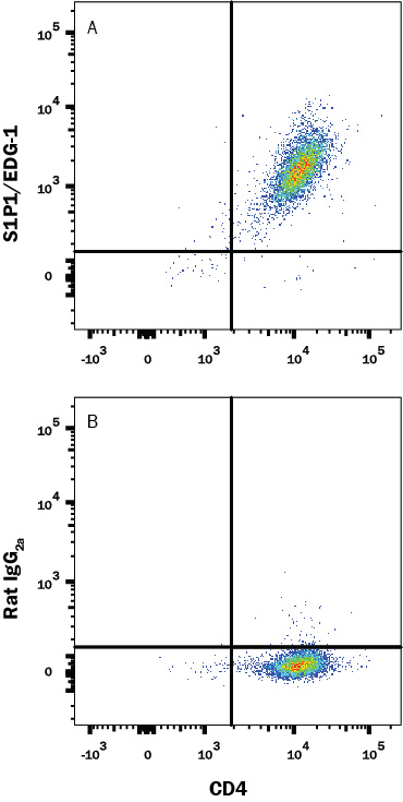
Detection of S1P1/EDG‑1 in HEK293 Human Cell Line Transfected with Mouse S1P1/EDG-1 and eGFP by Flow Cytometry. HEK293 human embryonic kidney cell line transfected with mouse S1P1/EDG-1 and eGFP was stained with either (A) Rat Anti-Mouse S1P1/EDG-1 APC-conjugated Monoclonal Antibody (Catalog # FAB7089A) or (B) Rat IgG2AAllophycocyanin Isotype Control (Catalog # IC006A). View our protocol for Staining Membrane-associated Proteins.
 View Larger
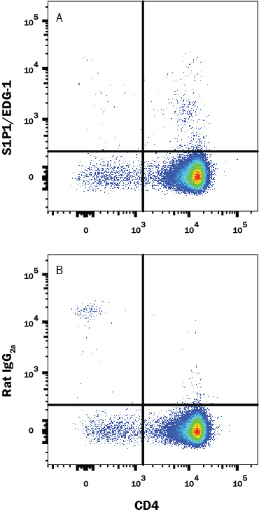
Detection of S1P1/EDG‑1 in Mouse Thymocytes by Flow Cytometry. Mouse thymocytes were stained with Rat Anti-Mouse CD4 PE-conjugated Monoclonal Antibody (Catalog # FAB554P) and either (A) Rat Anti-Mouse S1P1/EDG-1 APC-conjugated Monoclonal Antibody (Catalog # FAB7089A) or (B) Rat IgG2AAllophycocyanin Isotype Control (Catalog # IC006A). View our protocol for Staining Membrane-associated Proteins.
Reconstitution Calculator
Preparation and Storage
Background: S1P1/EDG-1
S1P1 (sphingosine 1-phosphate receptor-1), also known as EDG-1 (endothelial differentiation, G-protein coupled receptor-1) or S1PR1 (sphingosine-1-phosphate receptor 1) is a widely expressed, 37-40 kDa, G protein coupled receptor within the S1P subfamily of the EDG family. S1P1 is one of five receptors for the bioactive lipid S1P and mediates most of S1P effects on angiogenesis, vascular maturation, and cell migration, especially T cell egress from lymphoid organs. Human and mouse S1P1 share 84% amino acid identity within the N-terminal extracellular portion used as an immunogen.
Product Datasheets
Citations for Mouse S1P1/EDG-1 APC-conjugated Antibody
R&D Systems personnel manually curate a database that contains references using R&D Systems products. The data collected includes not only links to publications in PubMed, but also provides information about sample types, species, and experimental conditions.
6
Citations: Showing 1 - 6
Filter your results:
Filter by:
-
Targeting the MR1-MAIT cell axis improves vaccine efficacy and affords protection against viral pathogens
Authors: Rashu, R;Ninkov, M;Wardell, CM;Benoit, JM;Wang, NI;Meilleur, CE;D'Agostino, MR;Zhang, A;Feng, E;Saeedian, N;Bell, GI;Vahedi, F;Hess, DA;Barr, SD;Troyer, RM;Kang, CY;Ashkar, AA;Miller, MS;Haeryfar, SMM;
PLoS pathogens
Species: Mouse
Sample Types: Whole Cells
Applications: Flow Cytometry -
Sequestration of T cells in bone marrow in the setting of glioblastoma and other intracranial tumors
Authors: P Chongsathi, C Jackson, S Koyama, F Loebel, X Cui, SH Farber, K Woroniecka, AA Elsamadicy, CA Dechant, HR Kemeny, L Sanchez-Pe, TA Cheema, NC Souders, JE Herndon, JV Coumans, JI Everitt, BV Nahed, JH Sampson, MD Gunn, RL Martuza, G Dranoff, WT Curry, PE Fecci
Nat. Med., 2018-08-13;0(0):.
Species: Mouse
Sample Types: Whole Cells
Applications: Flow Cytometry -
Impaired lymphocyte trafficking in mice deficient in the kinase activity of PKN1
Authors: R Mashud, A Nomachi, A Hayakawa, K Kubouchi, S Danno, T Hirata, K Matsuo, T Nakayama, R Satoh, R Sugiura, M Abe, K Sakimura, S Wakana, H Ohsaki, S Kamoshida, H Mukai
Sci Rep, 2017-08-09;7(1):7663.
Species: Mouse
Sample Types: Whole Cells
Applications: Flow Cytometry -
IL4RA on lymphatic endothelial cells promotes T cell egress during sclerodermatous graft versus host disease
JCI Insight, 2016-08-04;1(12):.
Species: Mouse
Sample Types: Whole Cells
Applications: Flow Cytometry -
Targeting of Ly9 (CD229) Disrupts Marginal Zone and B1 B Cell Homeostasis and Antibody Responses.
Authors: Cuenca M, Romero X, Sintes J, Terhorst C, Engel P
J Immunol, 2015-12-14;196(2):726-37.
Species: Mouse
Sample Types: Whole Cells
Applications: Flow Cytometry -
Intravenous gammaglobulin inhibits encephalitogenic potential of pathogenic T cells and interferes with their trafficking to the central nervous system, implicating sphingosine-1 phosphate receptor 1-mammalian target of rapamycin axis.
Authors: Othy S, Hegde P, Topcu S, Sharma M, Maddur M, Lacroix-Desmazes S, Bayry J, Kaveri S
J Immunol, 2013-03-22;190(9):4535-41.
Species: Mouse
Sample Types: Whole Cells
Applications: Flow Cytometry
FAQs
-
Can staining with Mouse S1P1/EDG-1 conjugated Antibody, catalog # FAB7089*, be carried out at 4°C?
There is evidence that G protein family proteins require staining at room temperature (RT) or even 37 °C, so this is what we would recommend. Staining for 45 minutes is optimal with this antibody.
Reviews for Mouse S1P1/EDG-1 APC-conjugated Antibody
Average Rating: 4 (Based on 1 Review)
Have you used Mouse S1P1/EDG-1 APC-conjugated Antibody?
Submit a review and receive an Amazon gift card.
$25/€18/£15/$25CAN/¥75 Yuan/¥2500 Yen for a review with an image
$10/€7/£6/$10 CAD/¥70 Yuan/¥1110 Yen for a review without an image
Filter by:

